

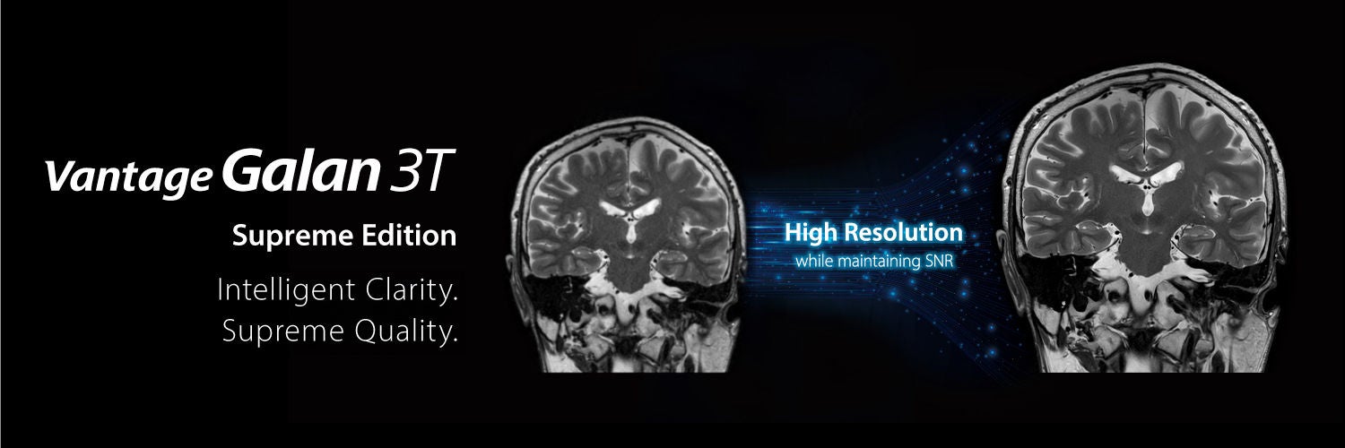
Supreme Image Quality
- PIQE delivers high SNR and high resolution for both 2D and 3D images to take MR imaging to the next level
- AiCE is applicable to a wide range of imaging including radial sampling pulse sequences
- Clear DWI images can be produced consistently with the combination of advanced averaging technologies, RDC DWI, Zoom DWI and PIQE
- Canon’s non-contrast MRA technology delivers consistent and non-invasive vascular imaging
Deep Learning Reconstruction
Precise IQ Engine (PIQE)
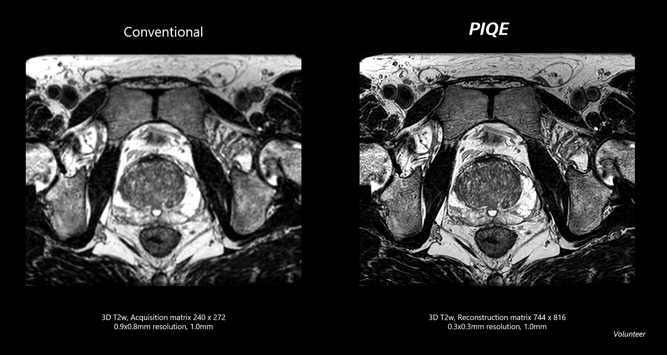
Precise IQ Engine (PIQE) is Canon’s high resolution Deep Learning Reconstruction for MRI. PIQE increases matrix size, removes noise, and delivers sharp anatomical images to take MR imaging to the next level.
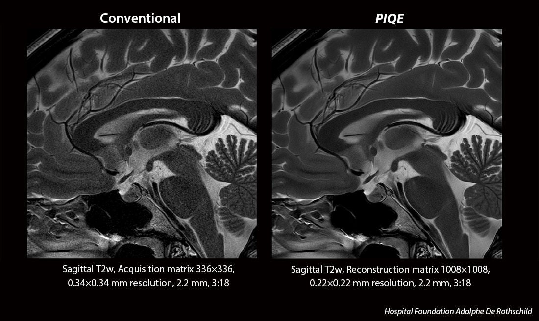
PIQE is now applicable to 3D sequences. High resolution 3D Image provides more detailed information.
Advanced intelligent Clear-IQ Engine (AiCE)
AiCE intelligently removes noise from images which results in higher SNR and enables increased resolution, as well as faster scan time when used in combination with unique accelerated scan applications.
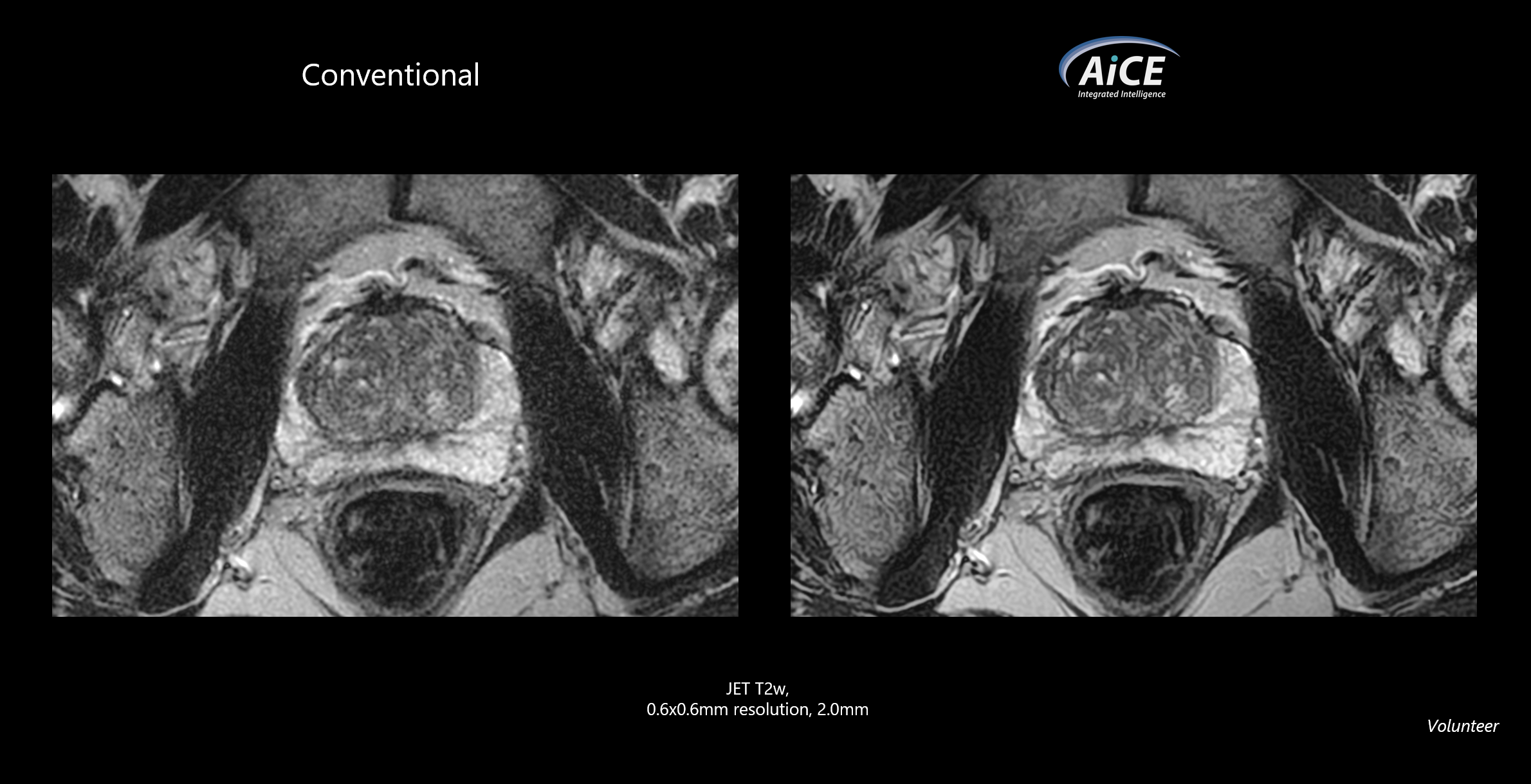
AiCE is applicable to a broad range of anatomies, contrast and applications, providing higher levels of imaging in every situation
All Contrast・Any Parameter・Across virtually All Sequences・Across a broad range of Anatomies・All RF coils
Now applicable to radial sampling pulse sequences
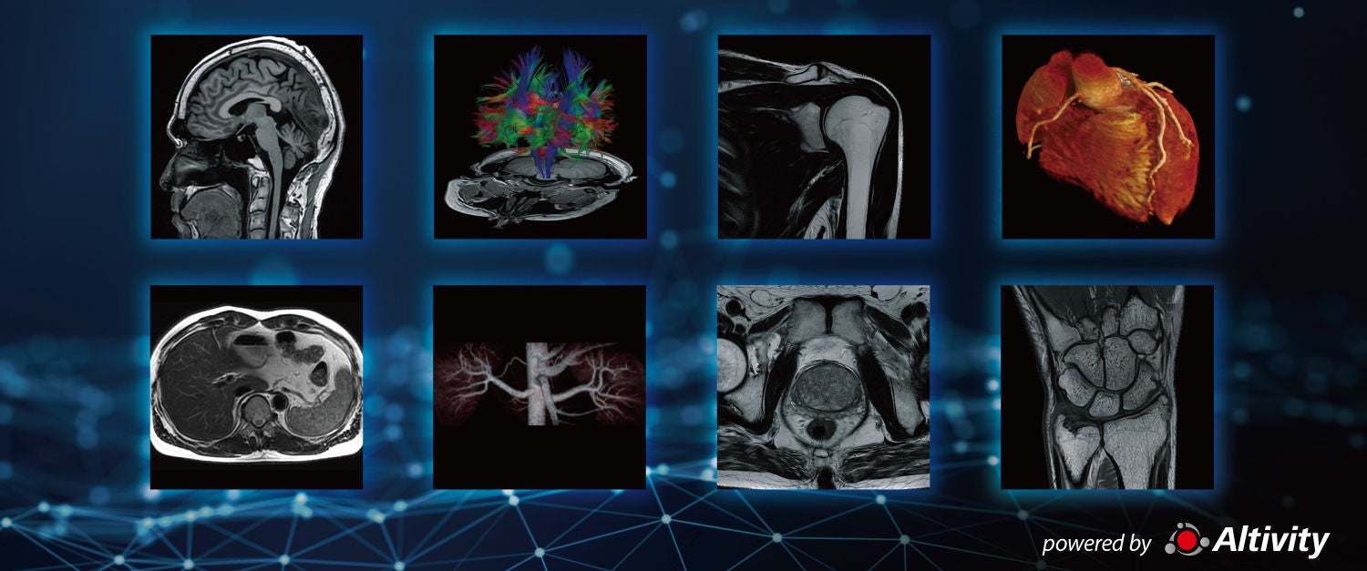
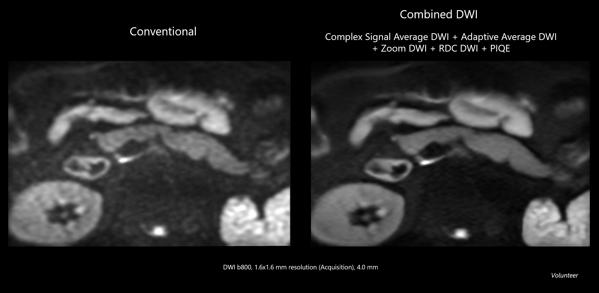
Combined DWI
Combined Intelligence delivers clear, consistent and confident DWI
By combining advanced signal averaging, distor tion control and resolution enhancement, our solution delivers stable, high-quality Diffusion Weighted Imaging across a wide range of clinical scenarios.
Complex Signal Average DWI
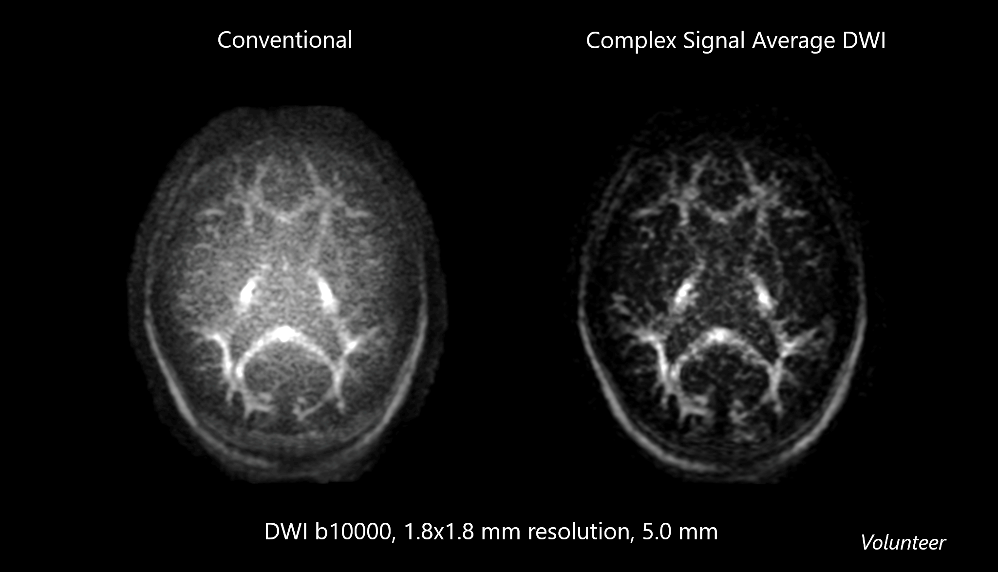
With conventional methods, noise signal is not able to be sufficiently suppressed even with an increased number of acquisitions (NAQ). Complex Signal Average uses Deep Learning–based phase correction to enable true complex-domain averaging.
By preserving the Gaussian noise characteristics of complex signals, it suppresses the background noise floor. Visualization of the signals typically dampened by noise is clearly enhanced compared to conventional magnitude averaging.
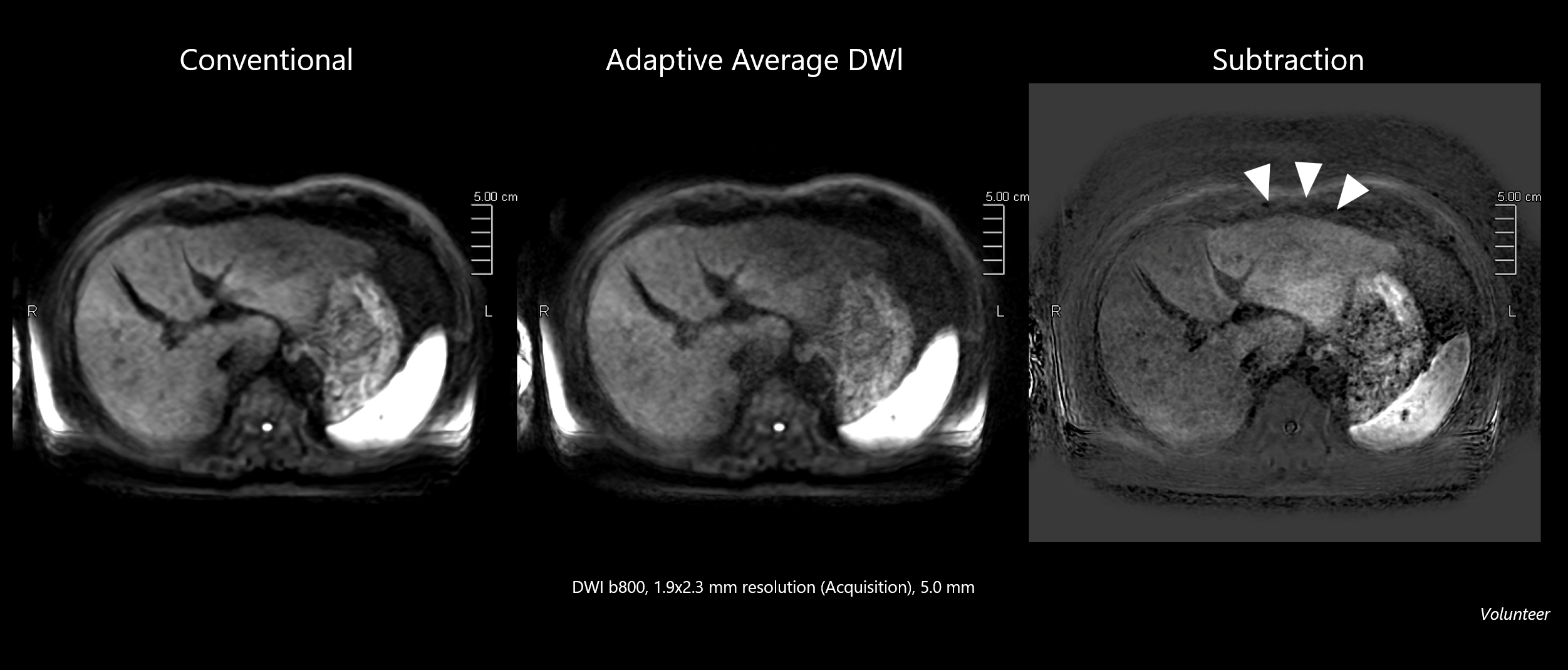
Adaptive Average DWI
In abdominal DWI, the signal intensity of the left lobe of the liver tends to be lower than that of the right lobe due to factors such as the timing of imaging and the influence of cardiac motion.
With this method, by weighting the signal intensity for each addition, it is expected that the reduced signal intensity of the left lobe of the liver will be improved.
RDC DWI
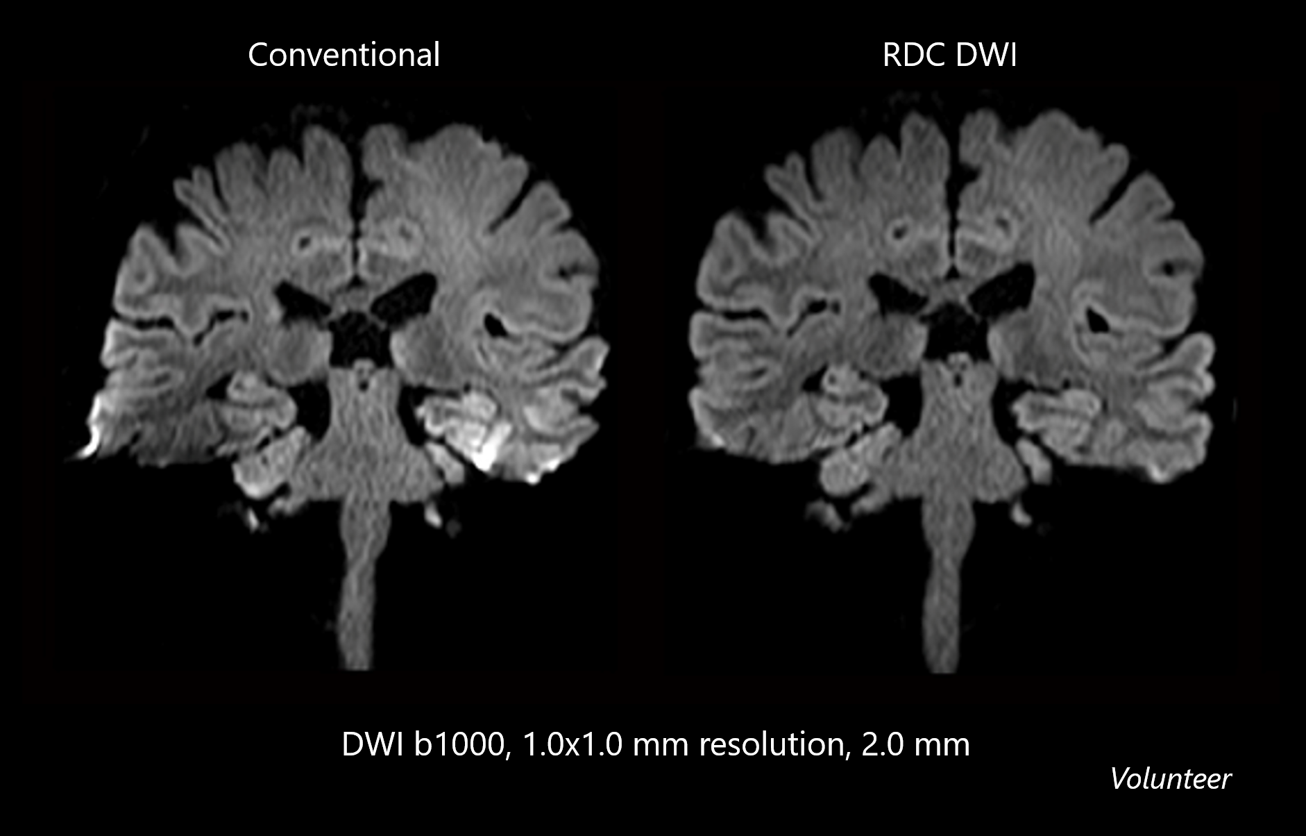
RDC DWI (Reverse encoding Distortion Correction DWI) is intended to reduce distortion in phase encoding direction due to B0 field inhomogeneity, or eddy current, in DWI sequence.
Zoom DWI
Diffusion weighted image with small FOV can be acquired suppressing distortion and unfolding errors, diagnostic reliability is expanded.
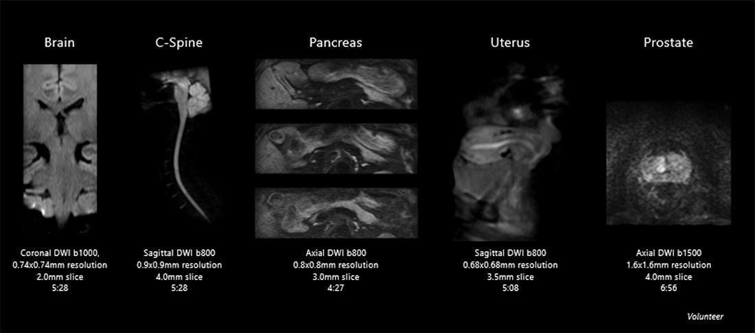
Non-Contrast MR Angiography
Vascular and blood flow assessment in all areas is performed without the use of contrast media.
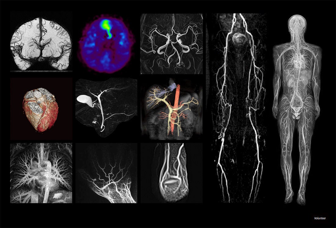
UTE MRA with CG Recon
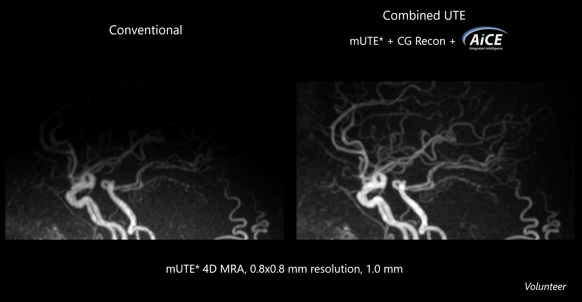 *mUTE: minimized acoustic noise utilizing UTE
Ultra-short TE (UTE) sequence enables the ability to visualize regions with short TE like lung, bone and tendon, as well as reducing artifacts caused by metallic devices in MR Angiography. AiCE reduces noise while Conjugate Gradient Reconstruction (CG Recon) enhances the accuracy of signal estimation and realizes more detailed visualization of the tissue or vasculature.
Enhanced multi-echo Flow Insensitive Black Blood (emFIBB)
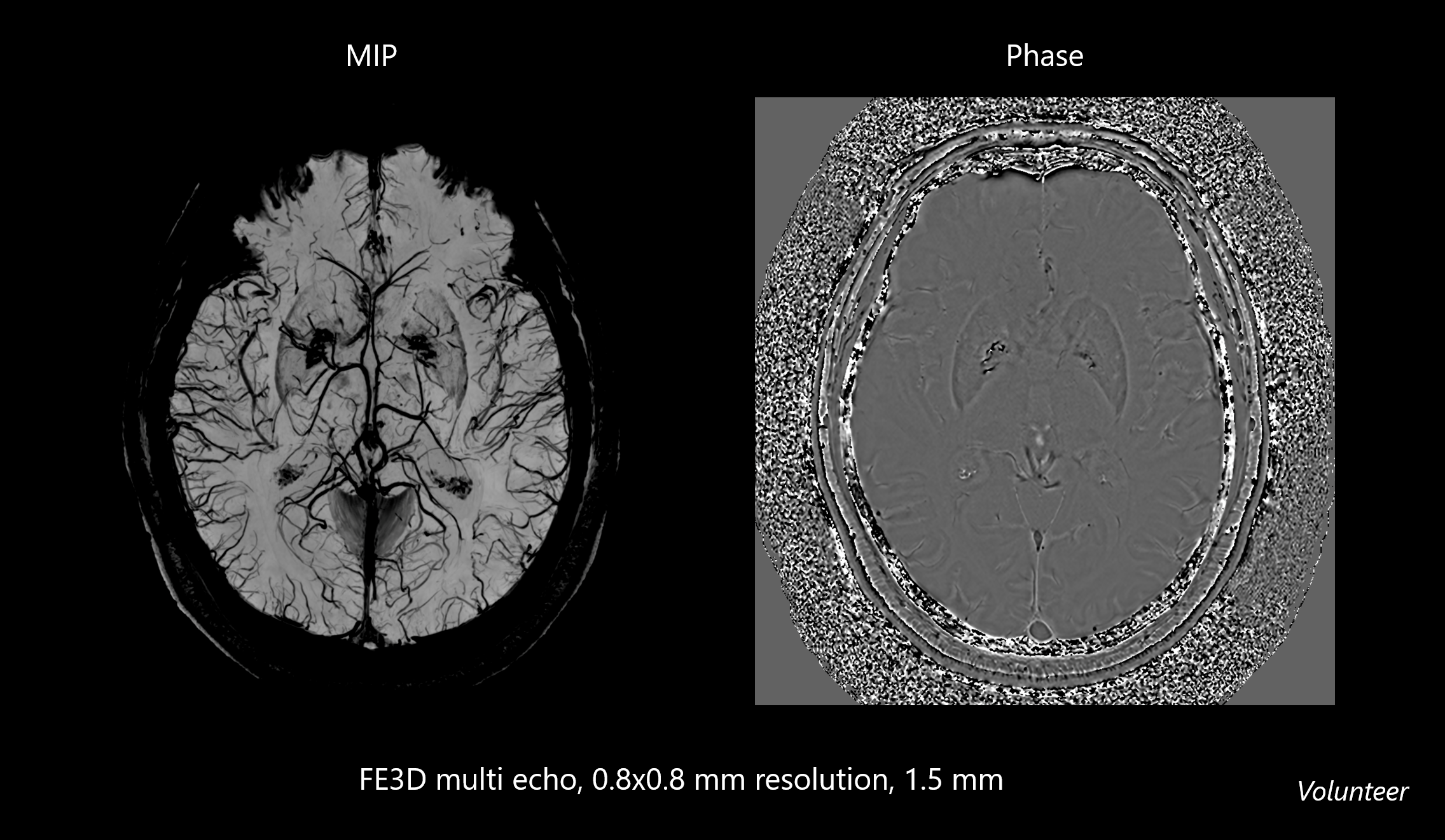
emFIBB has made it possible to optimize TE, TR and echo numbers enhanced by cosine filter. This results in enhanced SNR and contrast flexibility. With this method, arteries are clearly visualized with minimal interference from veins.
Pre-pulsed WFS
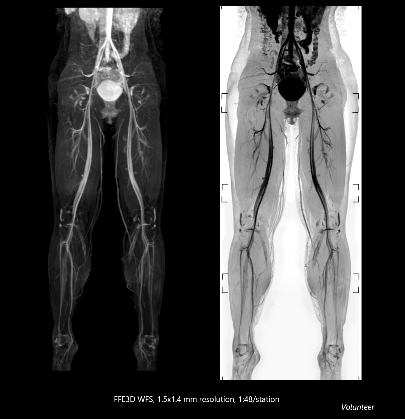
By enabling advanced preparation pulses combined with Water-Fat Separation (WFS), MRI contrast design is significantly expanded beyond conventional fat suppression. This approach delivers robust and uniform contrast while minimizing fat-related artifacts, and also enables non-contrast MRA to be acquired in a short scan time, reducing workflow complexity and increasing patient comfort.
4D Flow imaging
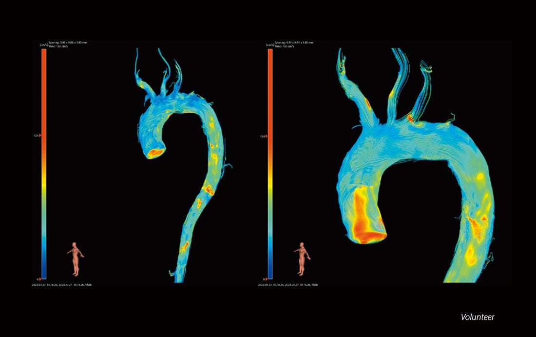
4D flow MRI offers the ability to measure and to visualize the temporal evolution of complex blood flow patterns within an acquired 3D volume.
Extended imaging range Improvement of Recon matrix with PIQE
- For 4D Flow imaging, analysis software is required. This image is using the software provided by Pie Medical Imaging.
Quantifiable MRI imaging to enhance diagnostic capability
Quantitative imaging techniques provide a wide range of options for referring physicians and staff. New techniques like MR Elastography and Fat Fraction Quantification (FFQ) for liver staging and quantification and contrast free Arterial Spin Labeling increase the imaging tools available for imaging various disease sets that were previously handled in other imaging modalities.
MR Elastography (MRE)
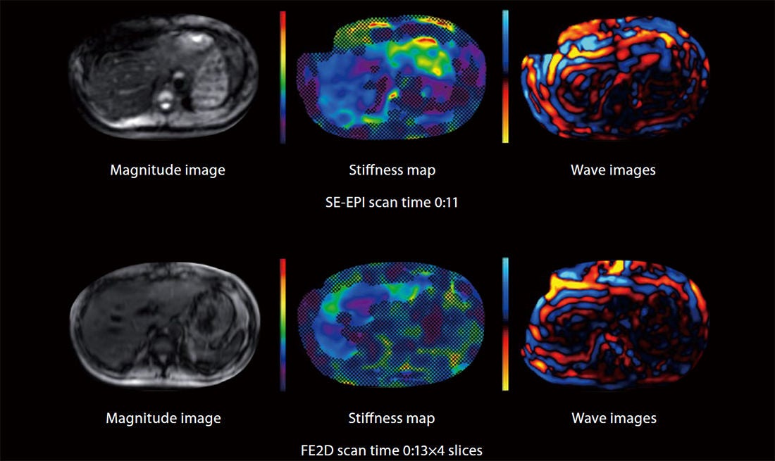
MRE is the only MRI technology that has been validated for staging liver fibrosis. The role of MRE has been increasingly recognized in multidisciplinary clinical guidelines for noninvasive liver fibrosis assessment, particularly in suspected cases of non-alcoholic fatty liver disease (NAFLD).
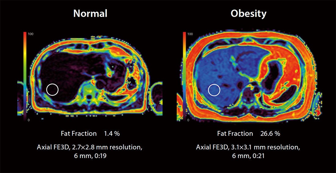
Non-invasive fat imaging and quantification
Imaging is rapidly becoming the standard for fat quantification. Canon’s fat imaging and quantification can simultaneously, in a single breath held exam, provide quantitative maps of the liver to measure proton density fat fraction (PDFF) and R2*.
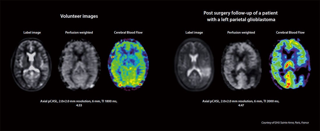
pseudo-Continuous Arterial Spin Labeling (pCASL)
Arterial Spin Labeling (ASL) MRI provides non-invasive methods to measure tissue perfusion without the use of external contrast agents. pCASL utilizes a fast spin echo (FSE) readout which makes it less sensitive to susceptibility artifacts and provides better image quality than other solutions.
Social Media


