
“Good to Know” Canon MRI
Canon’s Good to Know provides simple explanation to complex topic by our expert scientist team. Useful information is presented through handy and printable PDF, short videos and reference links.
Top 3 Articles
Topics

MPRAGE & MP2RAGE
Sara Pangaro, MS
WHAT?
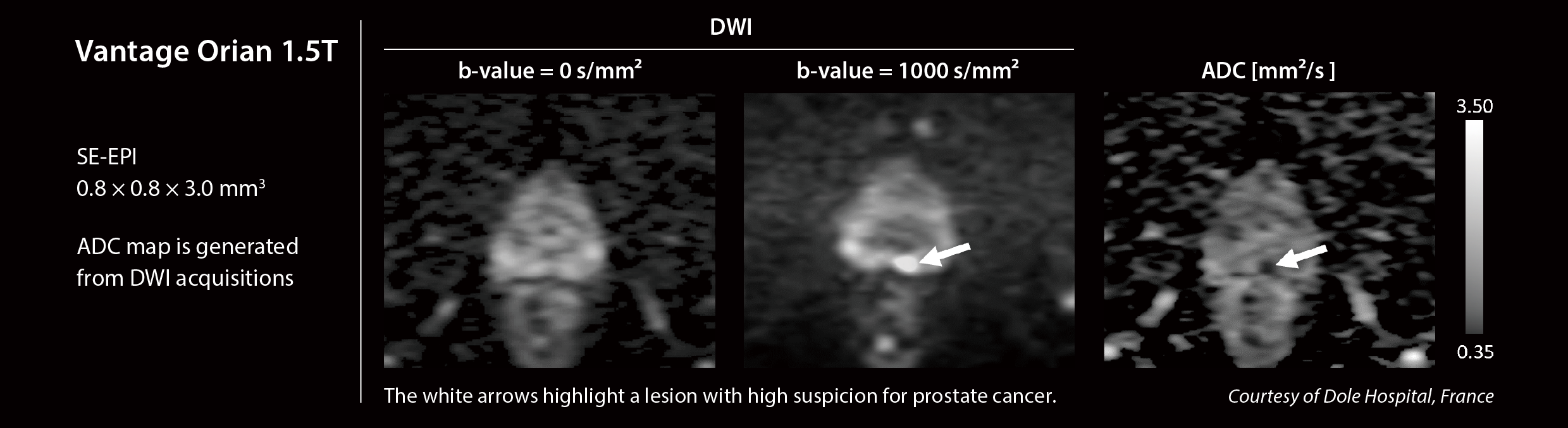
Processing of diffusion-weighted imaging (DWI) data to calculate Apparent Diffusion Coefficient (ADC) map.
WHY?
To quantify water diffusion within tissues and assess tissue microstructure.
WHEN?
Widely used in neuroimaging, oncology, and body imaging.
Download the full PDF for additional information (printable)
Internal links
White paper on ADC mapping applied to prostate cancers (by Dr. Tufik Bauab Jr., PhD)
Good to know on Computed Diffusion (by Thiele Kobus,PhD and Wolter de Graaf, PhD)
Good to Know on Multi-b Diffusion (by Alicia Palomar)
External links (may require logins)
Simple explanation of DTI by imaios.com
Simple explanation on stroke diagnosis and imaging by stroke-manual.com
Peer-reviewed paper on DWI for prostate cancer (Eur J Radiol. 2023)
Peer-reviewed paper on multi parametric MRI for pancreas imaging in diabetic patients (Tomography 2025)
Peer-reviewed paper on DWI for head and neck tumors (Eur J Radiol. 2024)

T1 MOLLI
Chia-Ying Liu, PhD
WHAT?
A specialized pulse sequence modified from original Look Locker technique for myocardial T1 mapping.
WHY?
T1 measurement could be time consuming due to multiple data acquisition successively after magnetization inversion. MOLLI techniques enable a fast assessment of myocardial T1 in one breath hold.
WHEN?
Myocardial T1 mapping provides a non-invasive quantitative tool for tissue characterization in myocardial disease.
Download the full PDF for additional information (printable)

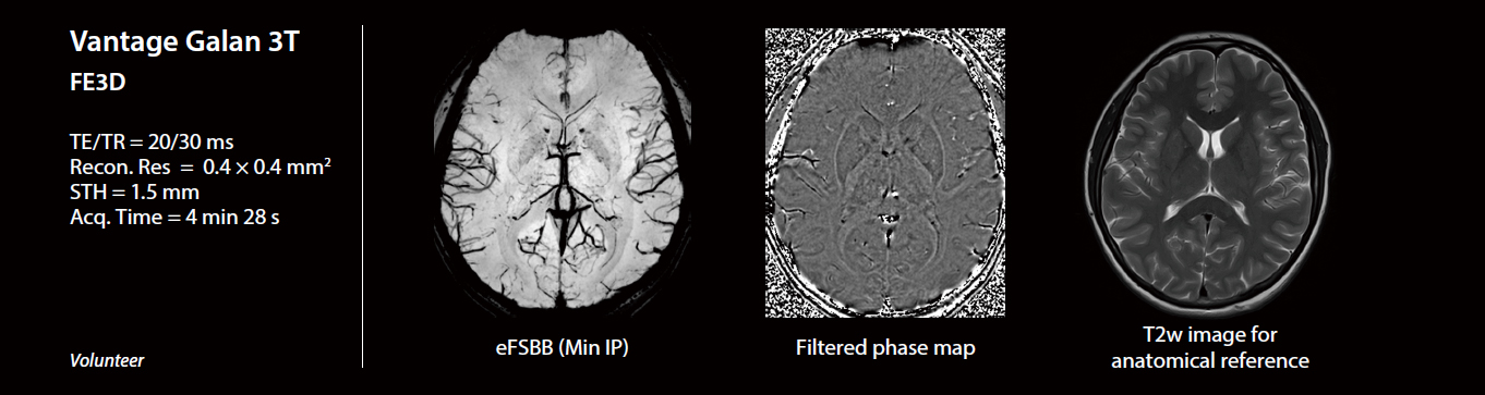
Flow Sensitive Black Blood (FSBB)
Alicia Palomar, MSc & MRes
WHAT?
Flow Sensitive Black Blood (FSBB) is sensitive to magnetic susceptibility differences between tissues.
WHY?
To exploit T2*-based dephasing effects and allow the visualization of different substances based on the magnetic field distortions that they produce.
WHEN?
For helping to assess local magnetic susceptibility changes present in multiple neurological conditions such as stroke, traumatic brain injuries, brain tumors, multiple sclerosis and neurodegenerative diseases.
Download the full PDF for additional information (printable)
Internal links
MR clinical case study of eFSBB for the detection of traumatic axonal injury
External links (may require logins)
Peer-reviewed paper describing the Cosine filter for obtaining eFSBB images (Magnetic Resonance in Medicine 2011)
Conference abstract evaluating the application of PIQE on FSBB imaging (ISMRM 2024)
Conference abstract on FSBB as a powerful technique in brain imaging (ECR 2025)
Simple definition of magnetic susceptibility by mriquestions.com
Simple explanation on susceptibility-weighted images acquisition and processing by mriquestions.com

Shape Coil
Erin J. Kelly, PhD
WHAT?
The Shape Coil is part of a new line of coil technology, offering flexibility in design for imaging many regions of the body including the torso, pelvis, joints, bones and extremities.
WHY?
The Shape Coil is flexible, soft, and light. It can be adapted to conform with each patient’s body shape and anatomy to improve patient comfort.
WHEN?
The Shape Coil can be used when more flexibility in positioning, orientation, acceleration, or coverage than traditional rigid or Flex coils is needed
Download the full PDF for additional information (printable)
Internal links
Shape Coil | MRI | Magnetic Resonance Imaging | Canon Medical Systems
External links (may require logins)
Magnetism - Questions and Answers in MRI (mriquestions.com)
RF coils: A practical guide for nonphysicists - PMC (nih.gov)

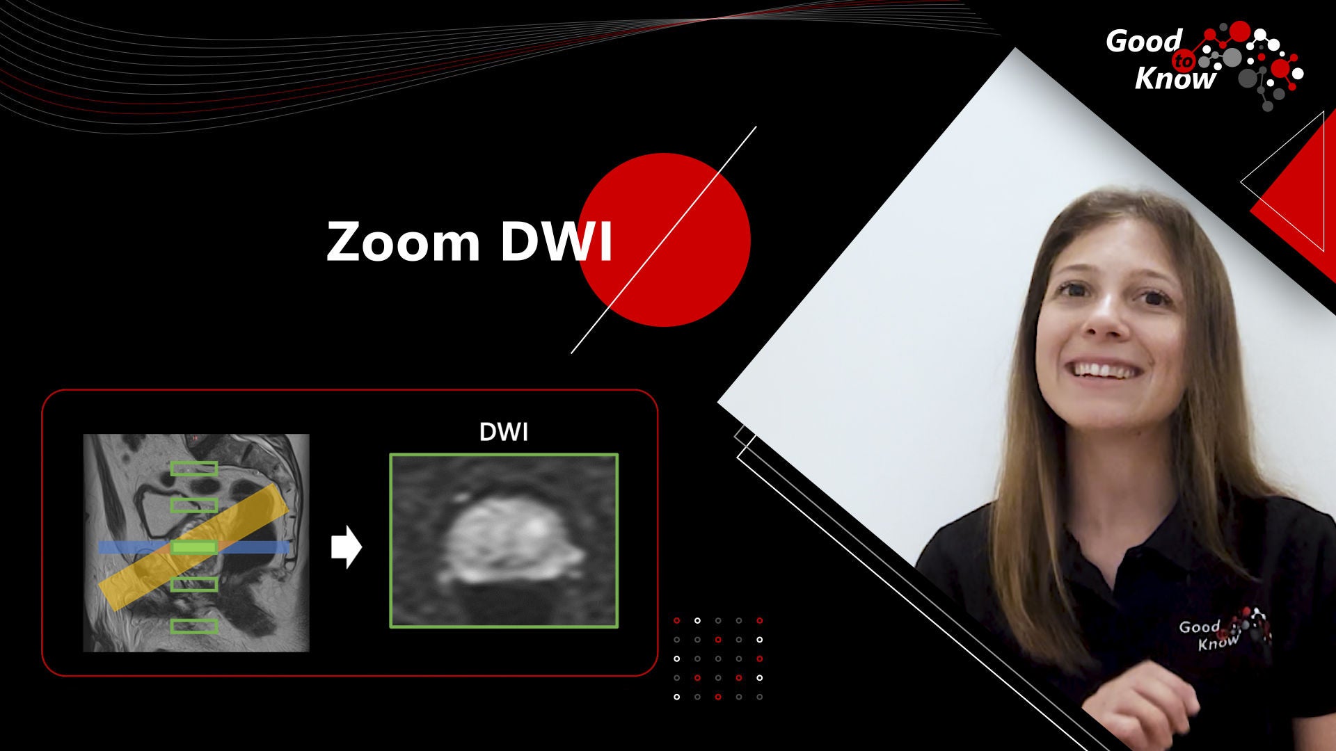
Zoom DWI
Wissam AlGhuraibawi, PhD
WHAT?
Zoom DWI is a technique that allows the acquisition of diffusion-weighted imaging (DWI) with reduced field of view (FOV), without aliasing, and with reduced blurring and geometrical distortions due to B0 inhomogeneity.
WHY?
In MR DWI, obtaining high-resolution images within a limited FOV is crucial when imaging small organs or tissue for better depiction of fine anatomical details and delineation of small lesions, such as tumors, infarcts, or other pathologies.
WHEN?
Zoom DWI is compatible with spin echo EPI sequences enabling high-resolution multi-slice DWI acquisitions in small FOV in the phase encoding (PE) direction such as in the brain, prostate, pancreas, breast, and C-spine.
Download the full PDF for additional information (printable)
Internal links
Technical explanations of ZOOM DWI technique (by Yuki Takai)
External links (may require logins)
Peer-reviewed paper evaluating ZOOM DTI on spinal cord patients (AJNR Am Neuroradiol. 2018)
Peer-reviewed paper evaluation reduced Field-of-View DWI on spinal cord patients (AJNR Am Neuroradiol. 2011)
Conjugate Gradient Reconstruction (CG Recon)
Chia-Ying Liu, PhD
WHAT?
CG Recon is a reconstruction technique that allows parallel imaging to be applied for non-Cartesian sampling acquisitions
WHY?
The aliasing that results from undersampled data is considerably more complex in non-Cartesian acquisitions and prevents straightforward unfolding by conventional parallel imaging methods
WHEN?
CG Recon enables parallel imaging for radial ultra-short echo time (UTE) imaging
Download the full PDF for additional information (printable)
Internal links
Good to Know on Parallel Imaging (by Min Xu, PhD)
External links (may require logins)
Review article on non-Cartesian parallel imaging reconstructions (JMRI 2014)
Peer-reviewed paper on Conjugate Gradient reconstruction applications (MRM 2001)
Conference abstract on 3D multi-echo UTE combined with CG Recon for CT-like imaging (ISMRM 2024)
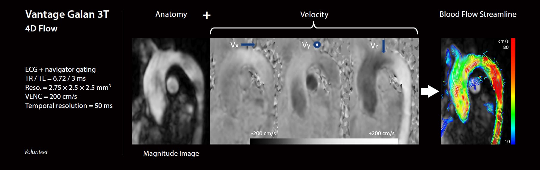
4D Flow Magnetic Resonance Imaging
Chia-Ying Liu, PhD
WHAT?
4D Flow refers to three-directional velocity encoding acquisition in a time-resolved manner.
WHY?
4D Flow provides capabilities for visualization and comprehensive analysis of complex blood flow patterns through any plane across the imaging volume.
WHEN?
To study both the qualitative and quantitative hemodynamic properties of blood flow for the diagnosis and risk-stratification of vascular abnormalities.
Download the full PDF for additional information (printable)
External links (may require logins)
Peer-reviewed article on 4D flow MRI consensus statement (JCMR 2023)
Peer-reviewed paper on 2D and 4D flow MRI comparison in patients with severe aortic stenosis (JCMR 2021)
Peer-reviewed paper on 4D flow MRI in patients with complex accelerated flow (JMRI 2013)
Peer-reviewed article on Phase Contrast imaging (RSNA 2020)
Simple explanations on PC-MRA by mriquestions.com
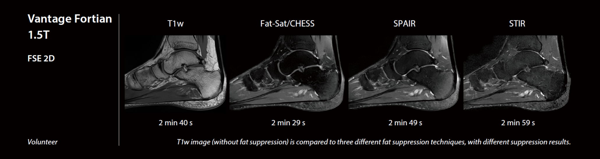
Fat Suppression Techniques
Alicia Palomar, MSc & MRes
WHAT?
Techniques to suppress the contribution of the adipose tissue from the total MRI signal.
WHY?
To improve contrast-to-noise ratio and the visibility of lesions, as well as to remove artifacts.
WHEN?
For multiple clinical applications (e.g. abdomen, breast and MSK imaging) and applicable to all MRI weightings.s.
Download the full PDF for additional information (printable)
IInternal links
Good to Know on the Dixon technique (by Min Xu)
White paper on Dixon efficiency for full spine and abdomen imaging (by Akihiko Arakawa)
External links (may require logins)
Peer-reviewed paper comparing STIR and CHESS Fat suppression for Time-SLIP renal MRA (Journal of Magnetic Resonance Imaging 2009)
Peer-reviewed paper evaluating Dixon as a sustitute of Fat-Sat for knee imaging (Journal of Medical Radiation Sciences 2019)
Peer-reviewed paper evaluating the effect of fat suppression on orbits, including CHESS and SPAIR techniques (Magnetic Resonance in Medical Sciences 2023)
Conference abstract on STIR pulse optimization for Time-SLIP renal MRA (ISMRM 2009)

Reverse encoding Distortion
Correction DWI (RDC DWI)
Angela Marin
WHAT?
Reverse encoding Distortion Correction (RDC) is a technique to reduce geometric distortion in Diffusion Weighted Imaging (DWI).
WHY?
The Echo Planar Imaging (EPI) technique, often used in DWI, is susceptible to distortions, especially in the phase encoding direction.
WHEN?
To reduce distortions in DWI while maintaining a short scan time.
Download the full PDF for additional information (printable)
External links (may require logins)
Simple explanations on Echo-planar Imaging (EPI) by mriquestions.com
Peer-reviewed paper on EPI fundamentals (Science 1991)
Peer-reviewed paper on EPI distortion correction (JMRI 2004)
Peer-reviewed paper on RDC DWI evaluation on phantom and healthy volunteers (Magn Reson Med Sci 2023)
Peer reviewed paper on RDC1 DWI applied to pituitary pathologies (Eur Rad Exp 2024)

Fast 3D Imaging
Niharika Gajawelli, PhD
WHAT?
Advanced approach to reduce 3D imaging scan time by utilizing efficient k-space sampling methods.
WHY?
To acquire 3D images faster, with up to about 50 % reduction in the scan time.
WHEN?
Applicable when acquiring 3D volumes in various anatomies. Some examples include brain imaging or abdominal imaging where breath-holds are required.
Download the full PDF for additional information (printable)
Internal links
White paper on standardized 3D brain MR examination in 6 minutes (by Vincent Dousset, MD PhD)
White paper on high resolution 3D free breathing technique (by Kohei Hamamoto, MD)
External links (may require logins)
Peer-reviewed paper on Fast 3D for breath-hold 3D magnetic resonance cholangiopancreatography (Eur J Radiol 2021)
Peer-reviewed paper on Fast 3D and AiCE for non-contrast MR coronary angiography (Can Assoc Radiol J 2021)
Conference abstract on Fast 3D wheel for cerebral MR angiography (RSNA 2021)
Conference abstract on fast 3D techniques to reduce scan time (RSNA 2021)
Iterative Motion Correction (IMC)
Wissam AlGhuraibawi, PhD
WHAT?
IMC is a Deep Learning-based motion correction method to improve the quality of images affected by patient motion.
WHY?
Patient motion creates artifacts that can cause images to lose their diagnostic value and may necessitate a repeated scan.
WHEN?
IMC can reduce imaging artifacts from sporadic, rigid, and nonrigid patient motion in fast spin-echo (FSE) sequences. IMC supports multiple imaging weightings such as T2, T1 IR, STIR & FLAIR, and in any plane for brain and cervical spine examinations.
Download the full PDF for additional information (printable)
Internal links
White paper on IMC solution (by Srikant Kamesh Iyer, PhD)
External links (may require logins)
Conference abstract on IMC evaluation (ISMRM 2023)
Workshop abstract on shuffle encoding technique (ISMRM Workshop 2022)
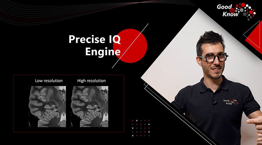
Precise IQ Engine (PIQE)
Valentin H. Prevost, PhD
WHAT?
PIQE is a solution designed to increase the reconstructed matrix size, providing finer resolution while maintaining or improving SNR.
WHY?
PIQE can lead to a higher throughput and/or a better clinical confidence.
WHEN?
PIQE can be used on 2D Fast Spin Echo (FSE) sequences, at both 1.5 and 3T, especially when SNR is low but more sharpness is needed.
Download the full PDF for additional information (printable)
Internal links
Good to Know on AiCE solution (by Hung Do, PhD)
White paper on PIQE testimonials (by MD and Daniel Chow, MD)
External links (may require logins)
Peer-reviewed paper on PIQE applied to MR angiography in patients (Neuroradiology 2023)
Peer-reviewed paper on PIQE applied to DWI in brain patients (Neuroradiology 2023)
Conference abstract on PIQE development (ISMRM 2023)
Conference abstract on PIQE clinical validation on knee patients (ISMRM 2023)

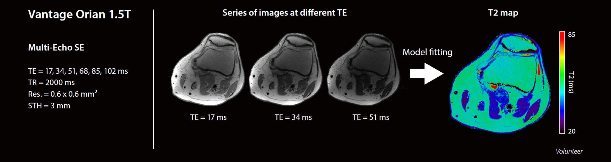
T2 Mapping
Iva Vilas-Boas Ribeiro, PhD
WHAT?
The standard method for T2 mapping is the multi-echo spin echo sequence to quantify transverse relaxation times (T2) in different tissues.
WHY?
T2 mapping quantitatively assesses tissue properties like water content and collagen structure, enabling objective analysis and enhancing detection of T2 relaxation time changes beyond qualitative signal intensity.
WHEN?
By quantifying T2 relaxation times across different tissues, this method is valuable in detecting abnormalities such as inflammation or fibrosis.
Download the full PDF for additional information (printable)
External links (may require logins)
Peer-reviewed paper on T1 and T2 mapping on pathological prostate (Acta Biomedica 2024)
Peer-reviewed paper on reference values for myocardial T2 mapping (JCMR 2015)
Review article on quantitative MSK imaging (American Journal of Roentgenology, 2019)
Review article on quantitative cardiac imaging (Frontiers in Cardiovascular Medicine, 2022)

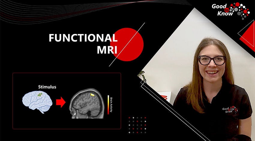
Functional MRI (fMRI)
Alicia Palomar, MSc & MRes
WHAT?
Fast scans of the whole brain every few seconds along a rest-task paradigm or during resting state.
WHY?
To assess neural activity based on the detection of blood oxygen level dependent (BOLD) changes.
WHEN?
For multiple purposes such as functional area mapping, evaluation of brain state (related to stroke, trauma or neurodegenerative disorders) and pre-surgical planning.
Download the full PDF for additional information (printable)
External links (may require logins)
Simple explanations on fMRI by mriquestions.com
Recommendations regarding fMRI paradigms (ASFNR website)
Wikipedia webpage of QIBA profiles, including fMRI for sensorimotor mapping
Peer-reviewed paper on aging biomarkers, including rs-fMRI (Mech Ageing Dev. 2020)
Task-fMRI part of the Human connectome project

ADC Mapping
Daisy Villano, MSc
Olea Medical
Olea Medical
WHAT?
Two specialized pulse sequences, FFE 3D MPRAGE and MP2RAGE, used in research and clinical settings for neuro-imaging.
WHY?
MPRAGE enables the acquisition of images with high SNR, fast scan time, and excellent tissue contrast.
MP2RAGE enables the detection and characterization of abnormalities in different conditions such as neurodegenerative diseases.
WHEN?
MPRAGE is used for most clinical applications, and it is suitable for advanced post-processing applications such as segmentation.
MP2RAGE is used for T1 mapping and for tissue characterization.

Download the full PDF for additional information (printable)
External links (may require logins)
Peer-reviewed paper on MP2RAGE fundamentals (NeuroImage 2010)
Peer-reviewed paper on T1 metrics preservation after AiCE denoising (Journal of Neuroradiology 2024)
Peer-reviewed paper on T1 mapping of hip cartilage using MP2RAGE (Radiology 2021)
Peer-reviewed paper on aging biomarkers, including MPRAGE (Mech Ageing Dev. 2020)
Peer-reviewed paper on FGATIR approach (also called WM-nulled) (NeuroImage 2009)
Metal Artifact Reduction
Erin J. Kelly, PhD
WHAT?
Metal artifact reduction strategies and sequences are designed to reduce susceptibility artifacts caused by the presence of metal in an MR imaging volume.
WHY?
In MRI, most metallic implants cause artifacts which can interfere with the anatomy of interest in the image by causing distortion or signal loss, due primarily to strong magnetic susceptibility.
WHEN?
In patients with metallic implants, artifact reduction techniques are especially useful for reducing the interference of related artifacts thus facilitating a more confident diagnosis.
Download the full PDF for additional information (printable)
Non-Contrast MR Angiography
Erin J. Kelly, PhD
WHAT?
A family of MR angiography techniques that utilize fresh spins to depict the contrast in blood vessels as an alternative to an injection of an exogenous contrast agent. In particular, Fresh Blood Imaging (FBI), Flow Spoiled FBI (FS-FBI) and Time-Spatial Labeling Inversion Pulse (Time-SLIP), are discussed.
WHY?
Non-contrast angiography (NC-MRA) provides the safest option for MR Angiography. It can exquisitely depict arteries and veins throughout the body in 3 dimensions, while eliminating the need for gadolinium-based contrast agents.
WHEN?
Any time visualization of the arteries or veins is needed for clinical diagnosis and follow-up related to narrowing or blockage of blood vessels, NC-MRA can be used for evaluation. NC-MRA is especially important for those patients who have a contraindication to exogenous contrast agents.
Download the full PDF for additional information (printable)
Internal links
White paper on renal artery stenosis diagnosis (by Cesar H. Nomura, PhD)
White paper on fresh blood imaging for peripheral angiography (by Timothy Albert, MD)
White paper on Time-Spatial Labeling Inversion Pulse (by Timothy Albert, MD)
External links (may require logins)
Peer-reviewed paper on renal 3D MR angiography (MAGMA 2020)
Review article on Non-contrast enhanced MR angiography (JMRI 2012)
Peer-reviewed paper on portography with time-spatial labeling inversion pulses (JMRI 2009)
Multi-b Diffusion
Alicia Palomar, MSc & MRes
WHAT?
Acquisition of diffusion-weighted images (DWI) using multiple b-values.
WHY?
To improve sensitivity of DWI and obtain further information regarding tissue behavior (apart from pure diffusion).
WHEN?
For several clinical applications and all body regions, but mostly used for oncological purposes.
Download the full PDF for additional information (printable)
Magnetic Resonance Elastography
Wissam AlGhuraibawi, PhD
WHAT?
Elastography is an imaging technique that creates a map where the pixel value represents the stiffness of tissue (measured as the elastic modulus in kilopascals, kPa) at a given location.
WHY?
In many diseases, tissue stiffness can change. For example, stiffness increases with increasing fibrosis, which is directly linked to the clinical outcome of many diseases, such as liver cirrhosis, hepatitis, and steatohepatitis.
WHEN?
MRE is the most effective non-invasive technique for diagnosis, staging, disease monitoring, and treatment follow-up for multiple chronic liver diseases.
Download the full PDF for additional information (printable)
External links (may require logins)
Website of Resoundant (MRE Hardware provider)
Wikipedia webpage of QIBA profiles, including MR Elastography of the liver
Review article on MRE guidelines (Abdom Radiol 2022)
Review article on the MRE accuracy for staging liver fibrosis in patients (Clin Gastroenterol Hepatol 2015)
Review article on MRE, laboratory tests and ultrasound to detect fibrosis in patients (Hepatology 2017)

Ultra short TE multi-echo (UTE multi-echo)
Hung P. Do, PhD
WHAT?
An MRI pulse sequence that allows acquisition of multi-echo data with ultra short echo times (TEs).
WHY?
Ultra short TEs allows reception of signals from tissues with short T2* which can not be obtained with conventional field echo (gradient echo) sequences.
WHEN?
UTE multi-echo is available at both 1.5T and 3T and can be used to visualize tissues with short T2* and to generate associated T2* maps.
Download the full PDF for additional information (printable)
Advanced intelligent Clear-IQ Engine (AiCE)
Hung P. Do, PhD
WHAT?
A deep learning based reconstruction method for MRI that intelligently removes noise while maintaining feature integrity.
WHY?
To increase SNR of the reconstructed images. This increased SNR could be translated to increased resolution and/or shortened scan time. This could also enable high field-like image quality without high-field challenges (e.g. higher cost, B0 & B1 inhomogeneity, etc.).
WHEN?
Applicable to all anatomies and available at both 1.5T and 3T, for both wide and narrow bore system.
Download the full PDF for additional information (printable)
Internal links
White paper on knee exploration at 3T using AiCE (by Garry Gold, MD PhD)
White paper on full brain exploration at 3T using AiCE (by Vincent Dousset, MD PhD)
White paper on AiCE explanation (by Hung Do, PhD)
External links (may require logins)
Peer-reviewed paper on brain imaging at 3T using AiCE (Magn Reson Med Sci 2020)
Peer-reviewed paper on non-contrast coronary angiography at 3T using AiCE (Can Assoc Radiol J 2021)
Conference abstract on quantitative imaging on brain at 3T using AiCE (ISMRM 2020)
Conference abstract on multi-anatomy imaging at 1.5T using AiCE (ISMRM 2021)
Conference abstract on short scan time imaging at 1.5T using AiCE (ISMRM 2021)
Arterial Spin Labeling
Valentin H. Prevost, PhD
WHAT?
Arterial Spin Labeling (ASL) method allows quantitative imaging of blood flow perfusion without the use of external contrast agent.
WHY?
Non-contrast angiography (NC-MRA) provides the safest option for MR Angiography. It can exquisitely depict arteries and veins throughout the body in 3 dimensions, while eliminating the need for gadolinium-based contrast agents.
WHEN?
To study perfusion disorders such as stroke or blood flow alterations induced by cancer, epilepsy, and neurodegenerative diseases.
Download the full PDF for additional information (printable)

Computed Diffusion (cDWI)
Thiele Kobus, PhD
Wolter de Graaf, PhD
Wolter de Graaf, PhD
WHAT?
Calculate new diffusion-weighted images from acquired images.
WHY?
To obtain additional diffusion information of the tissues without additional scan time.
WHEN?
Mostly used for oncologic purposes; high b-value images may result in better tumor conspicuity.
Download the full PDF for additional information (printable)

Compressed SPEEDER
Alba Iruela, MS
WHAT?
Acceleration technique based on incoherent undersampling in k-space.
It relies on the principles of compressed sensing technology.
WHY?
Compressed SPEEDER allows scan time reduction with higher acceleration factors than conventional SPEEDER while avoiding aliasing artifacts.
WHEN?
It is recommended for high-matrix situations, especially when the scanning conditions do not support traditional coil based parallel imaging.
Download the full PDF for additional information (printable)
Internal links
Good to Know on Parallel Imaging (by Min Xu, PhD)
White Paper on time reduction and additional benefits for MSK protocols (by Xavier Alomar, MD)
White Paper on workflow acceleration in neuro imaging (by Benoit Doche de Laquintane, MD)
White Paper on knee pathologies combining Compressed SPEEDER and AiCE (by Xavier Alomar, MD and Elena Ferre)
External links (may require logins)
Article on Compressed Sensing description and applications (IEEE Signal Processing Magazine 2008)
Review of Compressed Sensing from signal processing perspective (BMC Biomedical Engineering 2019)
Youtube Video on maths behind Compressed Sensing (by Steve Brunton, PhD)

Fat vs. Water, The Dixon Technique
Min Xu, PhD
WHAT?
One fat suppression approach based on the difference between the precessional frequencies of fat and water protons.
WHY?
To get a more efficient and reproducible fat signal suppression technique to improve visualization of lesions.
WHEN?
- For varied clinical applications, initially focused on abdominal regions, and then extended to musculoskeletal imaging, such as the neck, spine, the knee, brachial plexus, and the hands.
- Useful when conventional fat suppression technique is not reliable (large FOV, neck and plexus, off-center imaging...)
Download the full PDF for additional information (printable)
Internal links
White paper on clinical application of Dixon technique on neck region (by Seppo Kortelainen, MD)
White paper on efficiency and usefulness of Dixon technique on the whole spine and the abdominal region (by Akihiko Arakawa, MD)
Good to Know on Fat Fraction Quantification (by Mo Kadbi, PhD)
External links (may require logins)
Peer-review paper on Dixon technique applied to knee exploration at 3T (J Med Radiat Sci 2019)
Simple explanations on Dixon technique by mriquestions.com
Review article on Dixon technique (JMR 2008)

Fat Fraction Quantification
Mo Kadbi, PhD
WHAT?
A single breath-hold multi-echo Field Echo scan to accurately and reliably measure Proton Density Fat Fraction (PDFF) and R2*, even in the presence of increased iron concentration.
WHY?
To simultaneously provide, with one scan, quantitative maps of liver fat and R2*, in- & opposed-phase images, and fat- & water-only images.
WHEN?
Quantifying hepatic fat content and iron accumulation is needed for diagnosis, severity grading, disease monitoring, or treatment response assessment.
Download the full PDF for additional information (printable)
Internal links
Good to know on Dixon technique (by Min Xu, PhD)
External links (may require logins)
Educational video course on PDFF measurement (by Ali Piratsteh, MD)
Conference abstract on multi-center and multi-vendor PDFF quantification (ISMRM 2019)

Parallel Imaging SPEEDER and Exsper
Valentin H. Prevost, PhD
WHAT?
MRI images are created by gathering data from what is known in physics as the k-space. Parallel imaging (PI) under-samples data from the k-space by combining signal coming from multiple coils in parallel.
WHY?
By under-sampling data, we can speed-up scan time as less data is required in the acquisition phase.
WHEN?
Parallel Imaging can be used in several ways:
To go faster: higher patient throughput, shorter patient breath-hold, functional MRI, MR Angiography, reduce motion artifacts.
To reduce susceptibility artifacts : single shot EPI with Exsper(diffusion, DTI).
Download the full PDF for additional information (printable)

Social Media


