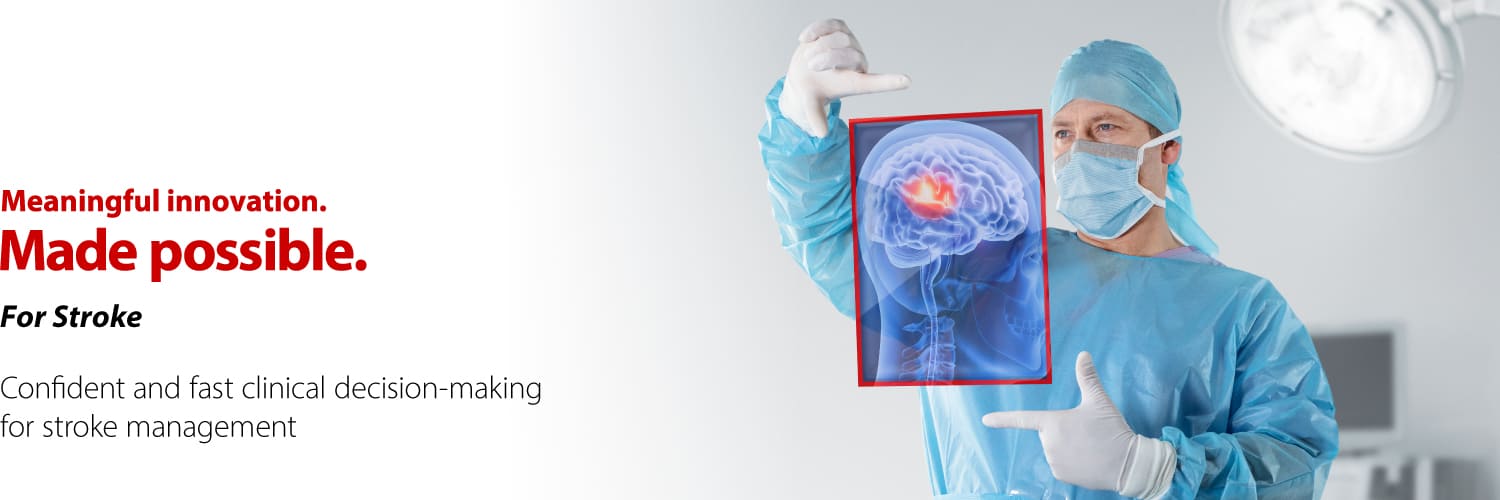
Stroke
Confidence where every second matters


Confident and fast clinical decision making across the patient pathway
At Canon Medical, we are committed to deliver innovative and advanced imaging solutions for confident and fast clinical decision making across the entire clinical pathway of stroke to support better patient outcomes. Together, we strive to minimize the burden of stroke on patients and society.

Our solutions
Our multi- modality, AI-enhanced solutions ensure seamless workflows when every second counts. The improved image quality and level of detail provided by our solutions allow for a more comprehensive understanding of the patient's condition which bolsters clinical confidence and decision-making.
Scanning fast without compromising image quality
Imaging in patients presenting with suspected acute stroke is an essential evaluation which should be done as fast as possible in order to establish the diagnosis, identify candidates for endovascular treatment and start the appropriate therapy without delays.
Our systems offer comprehensive examination of the brain and vessels with excellent AI-enhanced image quality in less than 5 minutes. Our unique deep learning reconstruction algorithm, Advanced intelligent Clear-IQ Engine (AiCE), has been designed to reduce noise and deliver sharp, clear images at speed.
Our CT solutions
In less than 5 minutes, a whole brain perfusion and 4D digital subtraction angiography of the intracranial circulation can be performed using our Aquilion ONE / INSIGHT Edition's 16-cm Area Detector CT.


Our MRI solutions
Our MRI Vantage series can support confirmation of recent onset of stroke and occluded blood vessels and associated lesions in less than 5 minutes.

Making decisions fast and confident
Optimized decision making with our AI solutions
In acute stroke, vast amounts of data needs to be interpreted immediately, so that the right treatment for each patient can be determined and implemented fast.
Automation Platform, an AI-driven solution, seamlessly streamlines your stroke workflow, from fast scanning to precise stroke triage and team notification, providing detailed clinical data to facilitate informed decision-making and ensure optimal stroke care. With CT brain perfusion imaging derived from Bayesian algorithm, you can now assess the extent of salvageable tissue, as well as indicate potential perfusion deficits to help diagnose and treat ischemic strokes.


Delivering therapy fast and safe
See more clearly with high-definition imaging
Intracranial vessels are small, tortuous and prone to dissection and rupture. The conventional flat panel detector technology, developed over two decades ago, is inadequate for the rapid evolving landscape of intracranial devices which are becoming finer, more intricate and complex.
Our Alphenix system provides an innovative technology with the world’s first Hi-Def detector—offering more than twice the spatial resolution of conventional flat panel detectors (FPD)—for resolving fine details. The Hi-Def detector enhances visualization during critical aspects of endovascular interventions and has the potential to translate into additional accuracy and precision in X-ray imaging guided procedures with enhanced real-time visualization during complex endovascular procedures.
Save time with multi-modality one room solution
Estimated Door-To-Recanalization Time < 30 mins.(5)

Patient transfer between multiple rooms and modalities can be a major cause of delays in treatment. Our one-room solution seamlessly integrates CT, ultrasound and angiography, delivering multi-disciplinary care with confidence.
No patient transfer is needed between multiple rooms, thereby reducing door-to-treatment time to within 30 mins (5), enhancing patient outcomes and increasing team productivity.

Stratify risk in more detail
Seeing the unseen with our ultrasound solutions
Unstable carotid artery plaques represent a significant, yet manageable, risk factor for ischemic stroke.
Our ultrasound technology facilitates physicians to detect and analyze carotid plaque, contributing to stroke risk evaluation and informing preventive measures for patients at elevated risk. It enables the immediate visualization of initial morphological and hemodynamic alterations in the carotid artery and intracranial vessels, which are precursors to cerebral stroke, through high-resolution imaging.
AI-enabled IMT-measurements and thorough B-mode plaque morphology assessment are covered by dedicated vascular probes supported by Canon’s intelligent Dynamic Micro-Slice (iDMS) active matrix technique.

Superb Micro-vascular Imaging (SMI) ultrasound technology visualizes vulnerable plaque biomarkers such as intraplaque microvascular flow signals.
Intraplaque neovascularization (IPN) plays a key role in plaque instability in patients with cerebrovascular disease. The unique SMI algorithm can show IPN without the need of invasive contrast injections.

“SMI may be a useful tool in bed-side assessment of IPN with a significant advantage over CEUS of not requiring an intravenous contrast injection, allowing for easier use in routine clinical practice.”
Dr. Mahtab Zamani, MD
Neurology Resident
Oslo University Hospital Rikshospitalet, Norway
General references
- World Stroke Organization (WSO): Global Stroke Fact Sheet 2022 https://www.world-stroke.org/assets/downloads/WSO_Global_Stroke_Fact_Sheet.pdf
- https://thestrokefoundation.org/disability-after-a-stroke/
- Saver, J. L.| Time is brain—quantified | Stroke (2006) https://www.ahajournals.org/doi/10.1161/01.str.0000196957.55928.ab
- Kunz, W. et al.|O-001Lifetime quality of life and cost consequences of treatment delays in endovascular thrombectomy for stroke based on hermes data | Journal of NeuroInterventional Surgery (2018) https://jnis.bmj.com/content/10/Suppl_2/A1
- JM Cappuzzo et al. | A Rapid Multidisciplinary Hybrid Approach to Stroke Care at University at Buffalo Neurosurgery | Endovascular Today (2023) https://evtoday.com/articles/2023-jan/a-rapid-multidisciplinary-hybrid-approach-to-stroke-care-at-university-at-buffalo-neurosurgery
Disclaimer
For the AI technology described in this website, deep learning technology is used in the design stage. The systems themselves do not have self-learning capabilities.
For more information on Altivity and Canon Medical Systems’ AI solutions, please visit here.

Social Media


