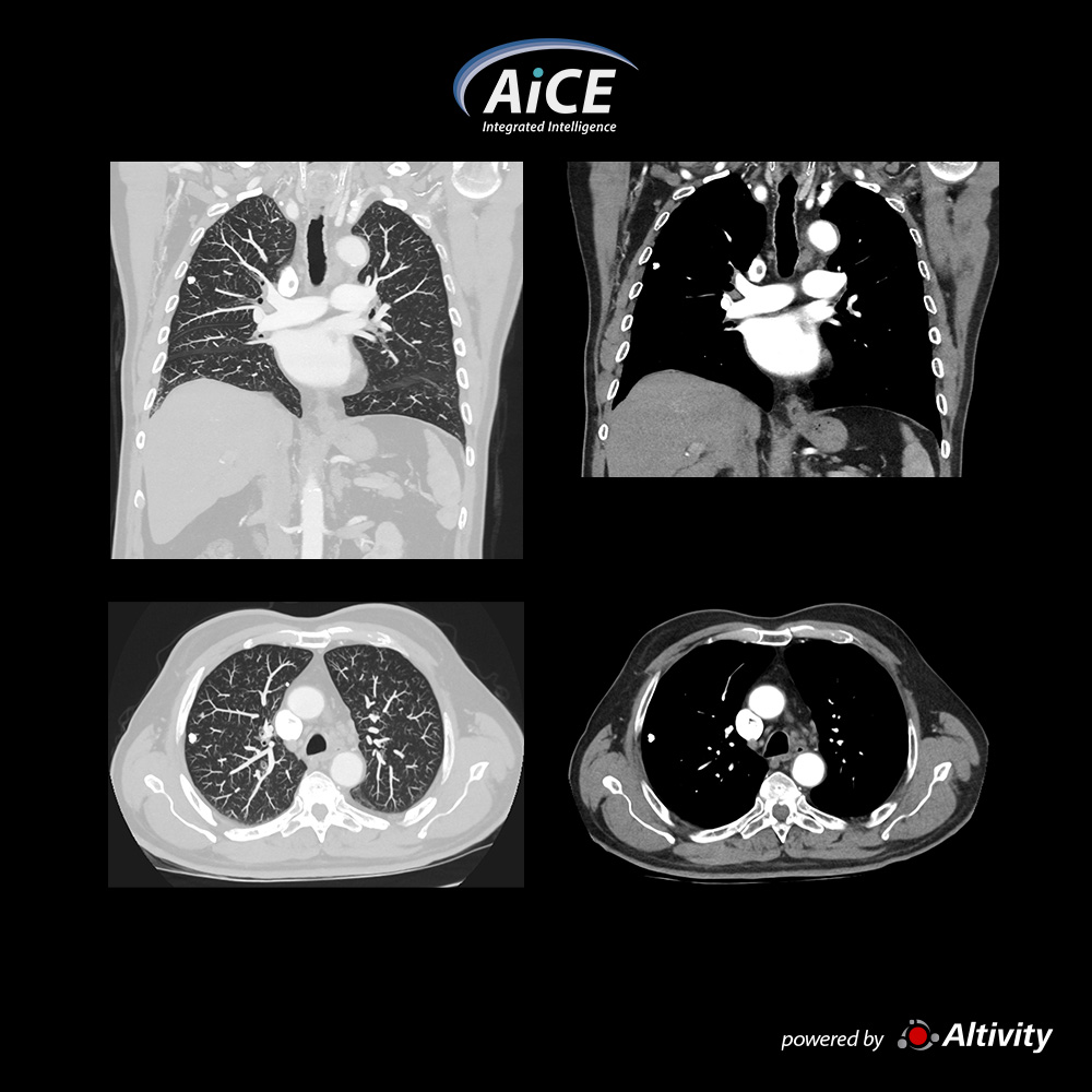
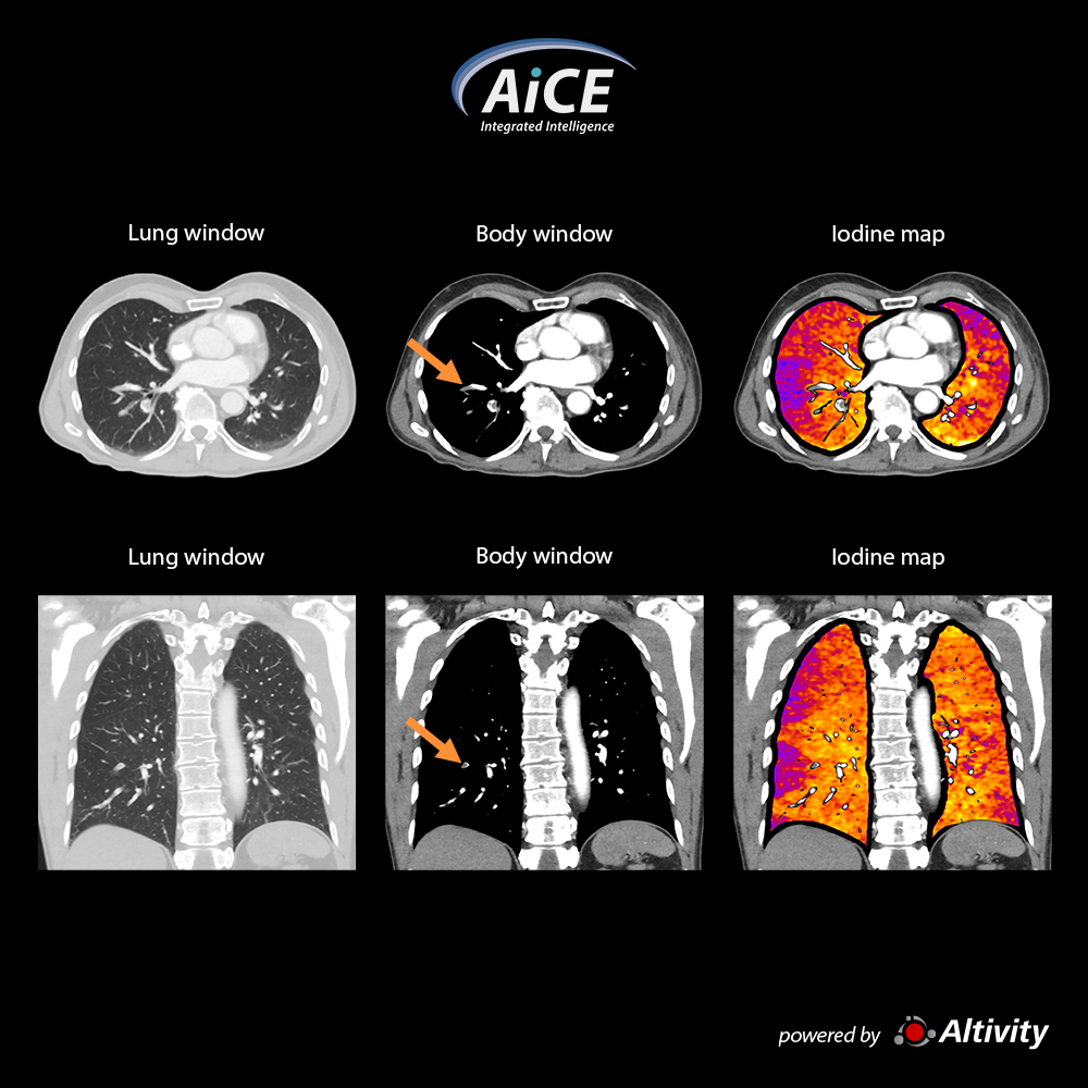
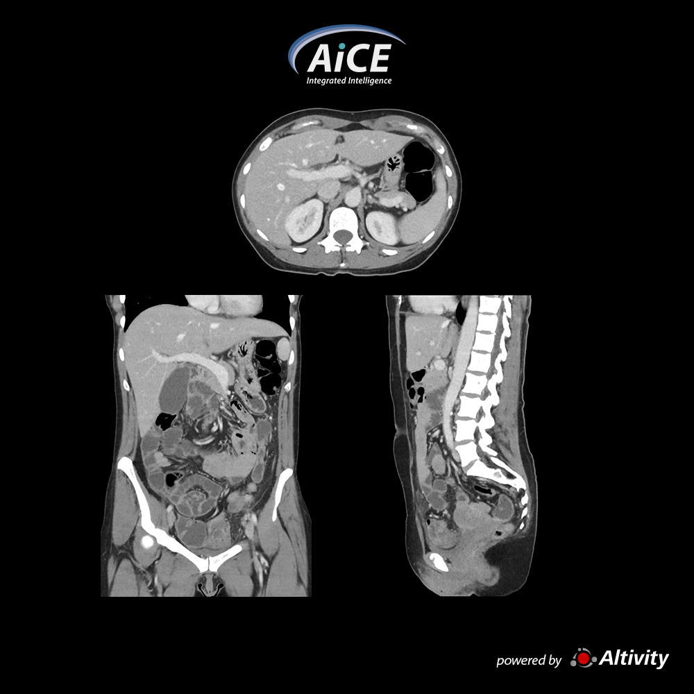
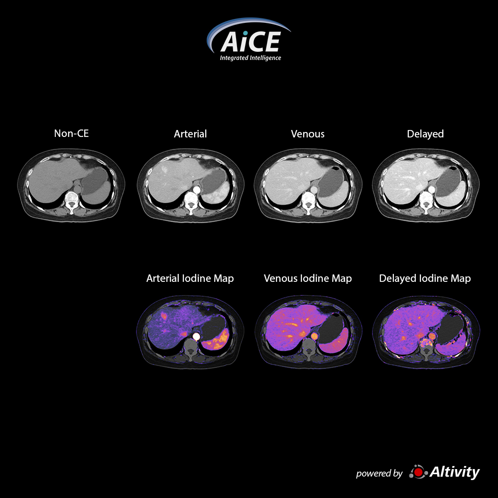
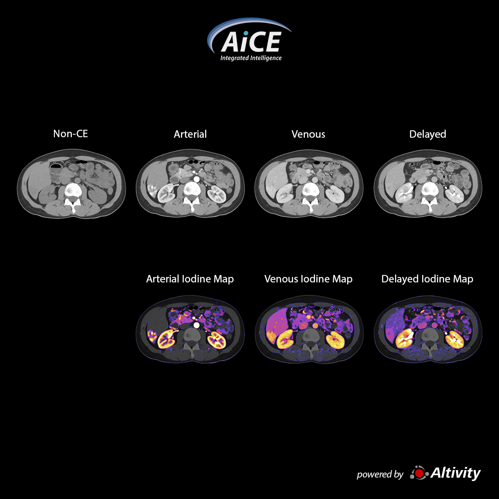
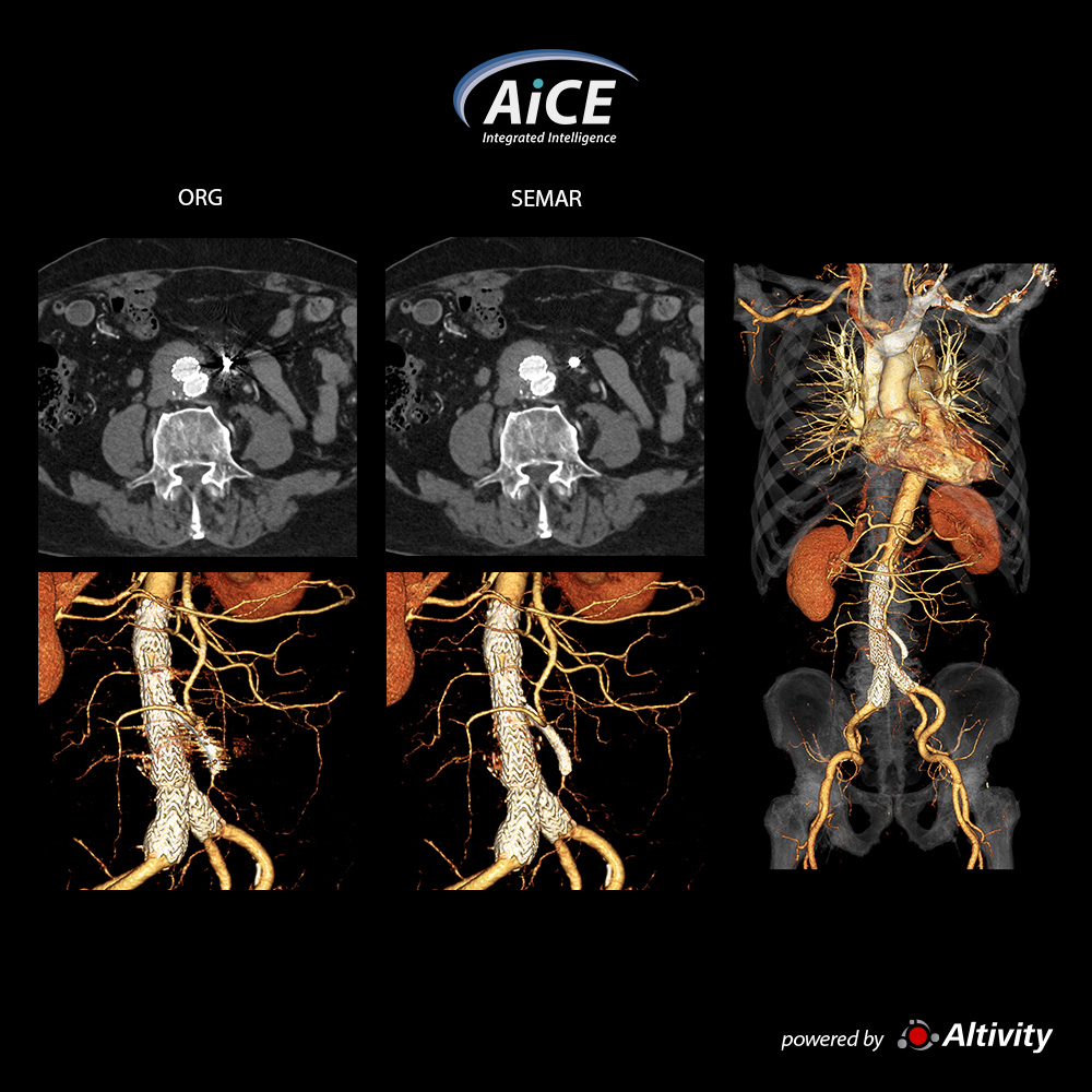
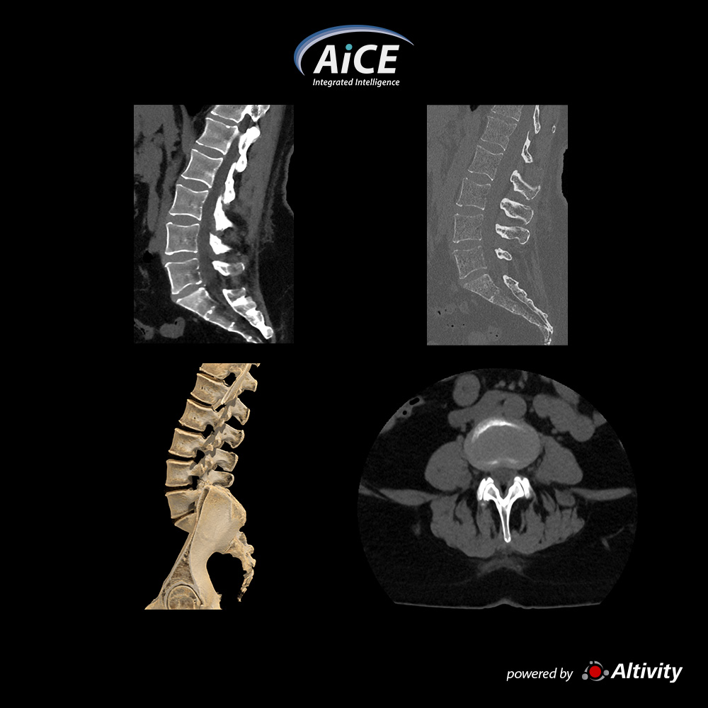
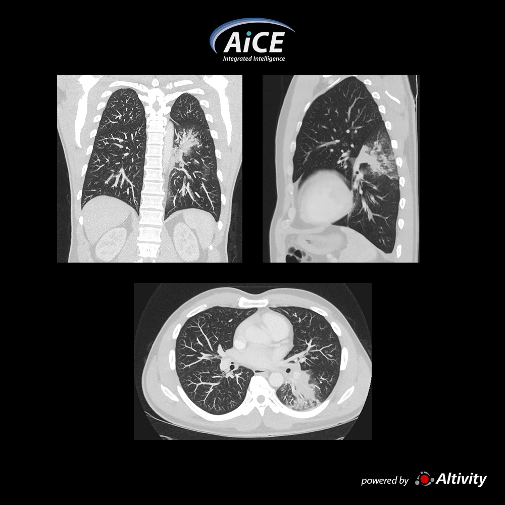
Chest - Pneumonia
Pneumonia seen in these MPR images of this 49-year-old man with shortness of breath and BMI 25.4
Courtesy
South Coast Radiology
Qld, Australia
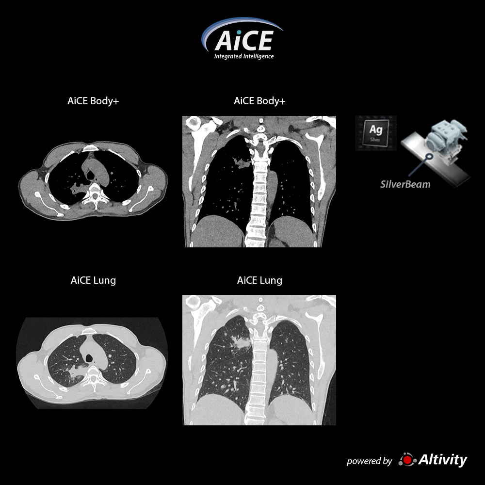
View Scan Parameters
Scan Mode
Ultra Helical
Collimation
0.5 mm × 80
kVp
120
mAs
SUREExposure
Rotation Time
0.35 s
Scan Range
409 mm
Dose Reduction
AiCE
CTDIvol
4.5 mGy
DLP
204.4 mGy·cm
Effective Dose
2.9 mSv
k-factor
0.014 (AAPM Report 96)
×
![拡大画像]()