

XR / White paper
How Much Does “AiCE ” Reduce Exposure in General Radiography?
Kazuya Mori
Department of Radiological Technology,
Saiseikai Kawaguchi General Hospital
Evidence-Based Radiographic Conditions for AiCE
At Saiseikai Kawaguchi General Hospital, there are three general radiography rooms for outpatients and inpatients. A total of 10 FPDs (CXDI) manufactured by Canon Inc. are used. The hospital began using “Advanced intelligent Clear-IQ Engine (AiCE)” in September 2023. This presentation introduces cases in which AiCE has improved image quality and reduced exposure dose at the hospital, and presents evidence to discuss an approach for determining appropriate radiographic conditions for AiCE.
Examples of application of AiCE at the hospital
AiCE is a noise reduction technology that uses deep learning, an AI technology, to train a neural network.
The key advantage of AiCE is that it resolves the problem in which images acquired using conventional noise reduction (cnvNR) become less clear due to part of the structural signal being removed together with noise, which results in decreased MTF. Furthermore, AiCE enables to provide less graininess in low-dose imaging by learning the noise characteristics specific to CXDI, significantly contributing to reducing exposure doses at the hospital.
Cases in which use of AiCE effectively improved image quality and reduced exposure dose are presented below.
Figure 1 shows a case of screw fixation from the cervical spine to the thoracic spine. In this case, the application of AiCE achieved a 28.6% dose reduction compared to cnvNR. In particular, in the shoulder region, which requires a high exposure dose in the lateral view, improved visualization of the vertebral endplate and the screw structure was observed, providing clearer visibility compared to cnvNR while achieving lower dose by AiCE.
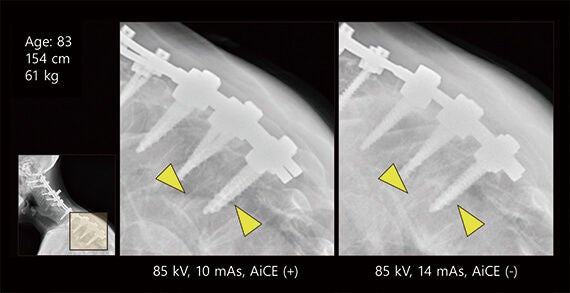
Figure 2 shows a case of L4-L5 lumbar spondylolisthesis in a 69-year-old male patient. Although the patient weighed 102 kg, the AiCE image clearly depicted the vertebral endplate even with a 38% reduction in exposure dose, clearly demonstrating the noise reduction effect.
Figure 3 shows a case of humeral fracture in a male infant. Some degree of callus formation can be observed in the AiCE image because the acquisition date of it differs from that of the image without AiCE applied. However, in the AiCE image, the fracture is clearly visible even with a 44% reduction in exposure dose.
Note that in extreme low-dose imaging, an insufficient dose may result in unclear structures, even though noise is reduced by AiCE. It is necessary to pay attention to this point when using AiCE. Appropriate exposure dose settings are required in order to maintain image quality, even when AiCE is used.
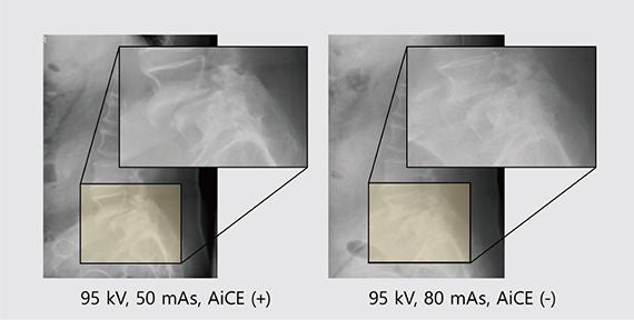
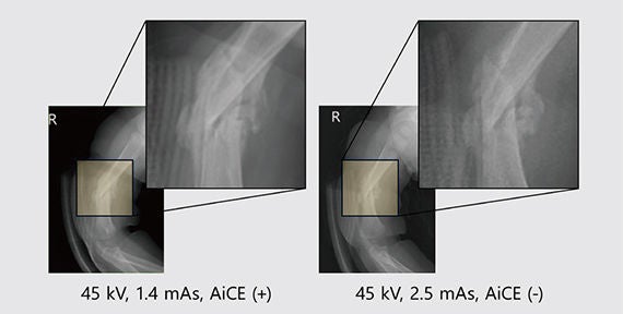
Figure 4 shows changes in exposure dose before and after the application of AiCE with consideration of appropriate exposure dose. With use of AiCE, approximately 25% to 35% dose reduction was achieved in X-ray imaging of the head, chest and abdomen, pediatric patients, vertebral bodies, and pelvis. Furthermore, when compared to the data in “National diagnostic reference levels in Japan (2025) - Japan DRLs 2025-”, approximately 25% to 50% dose reduction was achieved. For other regions such as the extremities, and for the head (frontal view), the hospital had already set the exposure dose to a lower level, and evaluation is performed with the dose reduction ratio maintained at approximately 20% with a margin of safety. Given the characteristics of AiCE, further dose reduction in the future is considered possible for these regions as well.
AiCE has proved to be effective in reducing noise in a variety of cases, including the extremities and pediatric imaging. In particular, in X-ray imaging for patients requiring a higher dose due to body thickness, high dose reduction effects have been achieved.
DRL2025 [mGy] | Ka,e [mGy] (cnvNR) | Ka,e [mGy] (AiCE) | Reduction ratio | Comparison with DRL2025 | |
| Head, frontal view | 1.3 | 1.39 | 1.04 | 25.35% | -20.20% |
| Chest, frontal view | 0.2 | 0.15 | 0.09 | 36.25% | -53.33% |
| Abdomen, frontal view | 1.4 | 1.43 | 0.91 | 36.11% | -34.96% |
| Infant chest, frontal view | 0.1 | 0.07 | 0.05 | 31.42% | -50.22% |
| Toddler chest, frontal view | 0.1 | 0.11 | 0.07 | 33.93% | -25.59% |
| Infant hip joint, frontal view | 0.1 | 0.08 | 0.06 | 30.93% | -43.72% |
| Cervical spine, frontal and lateral view | 0.5 | 0.59 | 0.39 | 33.58% | -22.19% |
| Thoracic spine, frontal view | 1.8 | 1.85 | 1.25 | 32.82% | -30.78% |
| Thoracic spine, lateral view | 3.4 | 3.27 | 2.24 | 31.51% | -34.21% |
| Lumbar spine, frontal view | 2.5 | 2.42 | 1.49 | 38.23% | -40.21% |
| Lumbar spine, lateral view | 5.5 | 5.49 | 3.50 | 36.28% | -36.34% |
| Pelvis, frontal view | 1.7 | 1.79 | 1.31 | 27.16% | -23.15% |
Appropriate radiographic conditions for AiCE
When the hospital first introduced AiCE, there were few papers on this new function, making it difficult to set radiographic conditions based on prior research.
Therefore, verifications using phantoms and evaluation of clinical images under various conditions were conducted at the hospital to determine appropriate radiographic conditions for AiCE.1)
1. Method
For evaluation of clinical images, we used Blind/Referenceless Image Spatial Quality Evaluator (BRISQUE), a method for Image Quality Assessment (IQA). BRISQUE is a machine learning-based image evaluation method in which lower scores relative to the magnitude of change indicate higher image quality.
To visually evaluate AiCE images, we used a chest phantom containing simulated tumors placed in the upper lung field and the mediastinum (behind the heart). Next, clinical images were retrospectively evaluated using the radiographic conditions obtained through the evaluation, and the validity of the radiographic conditions was verified. An image acquired at 2.4 mAs using AEC with cnvNR applied was used as the reference image, and images with dose reduced by 16% decrements (2.0 mAs, 1.6 mAs, 1.2 mAs, 0.8 mAs) were evaluated.
2. Subjects
The subjects were 100 patients who underwent both chest radiography using cnvNR and chest radiography using AiCE. The examinations were performed over a period of approximately six months, from September 2023 to March 2024. BRISQUE was used to evaluate image quality, and the entrance surface dose was measured to evaluate dose.
3. Results
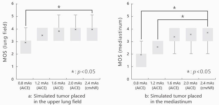
In the visual evaluation of the lung field, the AiCE image acquired at 0.8 mAs showed a significant decrease in visibility. However, the image acquired at 1.2 mAs, which is equivalent to half the dose of cnvNR, showed no significant difference in visibility, although the mean opinion score (MOS) decreased (Figure 5a). On the other hand, in the visual evaluation of the mediastinum, the image acquired at 1.2 mAs showed a significant decrease in the MOS value. Based on the above results, the hospital started clinical operation at 1.6 mAs, which is equivalent to a 32% dose reduction (Figure 5b).
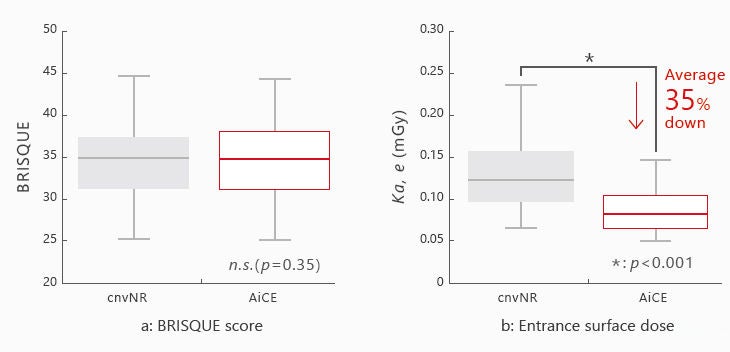
The results of the clinical image evaluation for cnvNR and AiCE (32% dose reduction) showed that cnvNR and AiCE were comparable in terms of the BRISQUE score (Figure 6a). In addition, when using AiCE, there was an approximately 35% reduction in the entrance surface doses compared to cnvNR (Figure 6b).
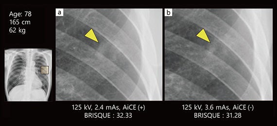
As an example of images actually used in clinical image evaluation, a case in which the dose was reduced by 33% is shown in Figure 7. In this case, a tumor is present in the left middle lung field, and there are no visually apparent changes resulting from dose reduction. Regarding the tumor area, the tumor border was clearly depicted in the AiCE image compared to the image using cnvNR.
To determine radiographic conditions, evaluation of images acquired using AiCE was conducted based on the phantom study using IQA. The results concluded that an average dose reduction of 35% is achievable compared to conventional radiographic conditions. The hospital plans to continue to expand the potential of the dose reduction effect of AiCE and further investigate the optimal dose by examining and evaluating radiographic conditions for each target region.
Conclusion
AiCE, a novel AI-based noise reduction processing technology, is a useful tool for easily acquiring low-dose, low-noise images without any disadvantages.
AiCE has the advantage of reducing invasiveness by reducing exposure dose during medical checkups or X-ray imaging for adult and pediatric patients. This is considered a significant benefit for medical institutions, regardless of facility size or type.
* The contents of this report include personal opinions of the author based on his clinical experience and knowledge.
* The AI technology described in this report is used in the design stage of the system. The system itself does not have self-learning capabilities.
* Also refer to the package insert for the medical device.
* Intelligent NR is a proprietary term of Canon Inc.
Advanced intelligent Clear-IQ Engine(AiCE) is the brand name that encompasses Intelligent NR.
* This article is based on the content of Luncheon Seminar 15 at the 41st Japan Conference of Radiological Technologists, titled “Evolution of Diagnostic Imaging Realized by Advanced Intelligent Clear-IQ Engine (AiCE): Next-generation Clinical Practice Led by Radiological Technologists Engaged in General Radiography and Mammography,” held on September 14 at the Courtyard by Marriott Fukui(Fukui City, Fukui Prefecture), co-hosted by Canon Medical Systems Corporation.
References
1) Mori, K., et al., Radiography, 31(3):102958, 2025.
https://doi.org/10.1016/j.radi.2025.102958
Kazuya Mori
2012 Graduated from Ibaraki Prefectural University of Health Sciences and joined the Department of Radiological Technology at Saiseikai Kawaguchi General Hospital.
Leader of the general radiography and angiography team of the department since 2022.
2024 Completed master’s program at the Graduate School of Human Health Sciences, Tokyo Metropolitan University.
Medical physicist and healthcare information technologist.
2026 Completed doctoral program at the Graduate School of Human Health Sciences, Tokyo Metropolitan University.
PhD (Radiological Science).
The content reflects information available at the time of publication and may differ from current information.
Social Media


