

Ultrasound / White paper
The role of ultrasound in muscle injuries in professional soccer

Introduction
Muscle injuries are among the most significant problems in professional soccer teams. They account for approximately 31% of all injuries that force players to stop playing or training. Typically, a squad of 25 players can expect around 15 muscle injuries per season.1, 2
Furthermore, they carry the highest injury burden of any injury type — defined as the number of days of absence per 1,000 hours of exposure, a measure that integrates both incidence and severity.2, 3
When a muscle injury occurs, the first question is invariably how long the player will be unavailable.
To answer it as quickly as possible, the medical staff activates a series of protocols aimed at identifying the type of injury and assessing its severity, in order to reach the most accurate, reliable and early diagnosis possible and thereby establish the prognosis. Within these protocols, imaging plays a fundamental role.
Two imaging modalities are currently used: magnetic resonance imaging (MRI) and ultrasound.
MRI is the gold standard for assessing most muscle injuries in elite athletes. Its high sensitivity for muscle edema and its specificity in identifying small tendinous and aponeurotic lesions make it the reference modality.
However, it is costly, takes longer to complete, is less comfortable for the athlete, and is not always rapidly available.5
Ultrasound, by contrast, has evolved considerably over the past decade, both in its resolution of superficial structures, where it surpasses that of MRI, and in the penetration and visualization of deeper structures. It is a safe and comfortable technique that allows the examiner to interact with the player and to perform dynamic maneuvers complementing the clinical examination.
Ultrasound devices are currently available at the training facilities of most professional clubs, and the examination can be repeated as often as necessary.
For these reasons, several authors and recent consensus statements recommend ultrasound as the first-line imaging modality for muscle injury, reserving MRI for severe injuries or when ultrasound findings are inconclusive.5, 6, 7
However, the real-world effectiveness of ultrasound is highly dependent on the examiner’s experience and on the type of injury, with small and deep lesions remaining difficult to assess even in the most experienced hands.7
In the medical department of Athletic Club, we consider ultrasound to play a central role in evaluating the injuries commonly sustained by our players. This role has grown progressively in recent years, to the point that we now regard the technique as indispensable in our daily practice.
Material
At the Athletic Club sports facilities, three ultrasound systems are installed adjacent to the training pitches: the new Aplio beyond, an Aplio i600 and an Aplio a. The Aplio beyond is equipped with the highperformance linear probes PLU-1005BT and PLU-1204BT as well as the PVU-674BT convex probe (Fig. 1). The Aplio i600 and Aplio a feature corresponding linear probes for musculoskeletal imaging.
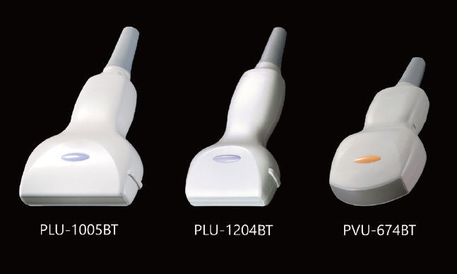 Fig. 1: Ultrasound probes currently used: Canon PLU-1005BT, PLU-1204BT and PVU-674BT
The PLU-1005BT probe is indispensable in musculoskeletal practice. It operates at frequencies from 3.8 to 14.5 MHz, with a center frequency of 10.0 MHz. It is a true workhorse probe, offering an excellent balance between penetration and resolution, with the added advantage of a field of view wider than the standard 5.8 cm. For these reasons, it is the ideal probe for assessing the most common muscle injuries, which occur in the thigh, leg and gluteal region.
The PLU-1204BT probe is highly recommended and operates at frequencies from 4.5 to 18.0 MHz. It is particularly at the upper end of this range that its greatest strength lies, providing excellent high-resolution images of superficial structures. This makes it especially useful for assessing the patellar tendon, Achilles tendon, ankle or foot ligaments, or for achieving superior definition of a specific lesion already detected with the PLU-1005BT.
The PVU-674BT probe is used far less frequently than the other two but has its specific utility. It is a small convex probe commonly used as a pediatric abdominal probe, operating at frequencies from 3.5 to 9.7 MHz. Its main advantage lies in providing deep penetration while maintaining good resolution. Additionally, being convex, it offers a wider field of view, making it ideal for studying very deep musculature such as the gluteal muscles, or for interventional procedures in the spine or hip.
Ultrasound and muscle injury
When evaluating a muscle injury with ultrasound, three findings should be systematically and comprehensively assessed: muscle edema, connective tissue lesions, and the presence of hemorrhage.
Muscle tissue injury
Skeletal muscle has a hierarchical structure worth reviewing (Fig. 2). The functional unit is the sarcomere, formed by the actin and myosin filaments that constitute the myofibrils. These group together to form cylindrical muscle cells (fibers), each surrounded by the endomysium; these fibers in turn group together to form fascicles.
Fascicles are surrounded by the perimysium, and the grouping of fascicles forms the muscle belly, which is enveloped by an outer layer known as the epimysium.9
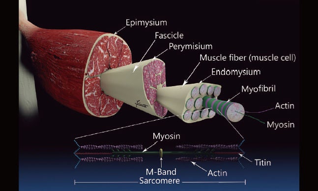 Fig. 2: Schematic representation of the hierarchical organization of the skeletal muscle architecture.
On axial ultrasound, normal muscle shows a starry-sky pattern (Fig. 3): a hypoechoic background corresponding to the muscle fibers themselves, with fine hyperechoic dots and lines representing the perimysium. These structures exhibit anisotropy, so that when the ultrasound beam is perpendicular to the fiber direction they appear more hyperechoic. In the longitudinal plane, the muscle displays a feathered or pennate pattern, with parallel hypoechoic bundles (muscle fibers) alternating with thinner hyperechoic ones (perimysium) (Fig. 4B).
When a muscle injury occurs, the ultrasound appearance changes: intramuscular edema and hemorrhage develop and, in more extensive tears, may be accompanied by fiber discontinuities filled with blood. Muscle edema appears as a hyperechoic haze that obscures some of the hyperechoic dots and lines of the perimysium (Figs. 4D, 5B, 6).
Adequate detection of muscle edema by ultrasound is technically demanding and requires adjusting the probe angle so that the beam is oriented perpendicular to the direction of the muscle fibers, which differs depending on the muscle being examined. For example, injuries to the aponeurotic expansion of the rectus femoris are located in the superficial and lateral portion of the muscle and are usually accompanied by significant muscle edema. Detecting this edema requires tilting the probe cranially to direct the ultrasound beam at an angle close to perpendicular to the fiber direction, thereby exploiting its anisotropy (Figs. 4B, 4D). Once the probe is correctly oriented, distal and proximal sweeps are performed, examining the muscle for alterations in the homogeneity of the muscular pattern and repeating these sweeps several times with small probe adjustments.
If this systematic approach is not followed, edema may go undetected.
Distinguishing anisotropy from edema is not always straightforward. It is important to bear in mind that edema delineates a distinct area relative to normal muscle, whereas anisotropy behaves uniformly across all muscle fibers sharing the same orientation. Furthermore, normal muscle preserves the hyperechoic images of the perimysium, whereas in edematous muscle these become blurred and disappear.
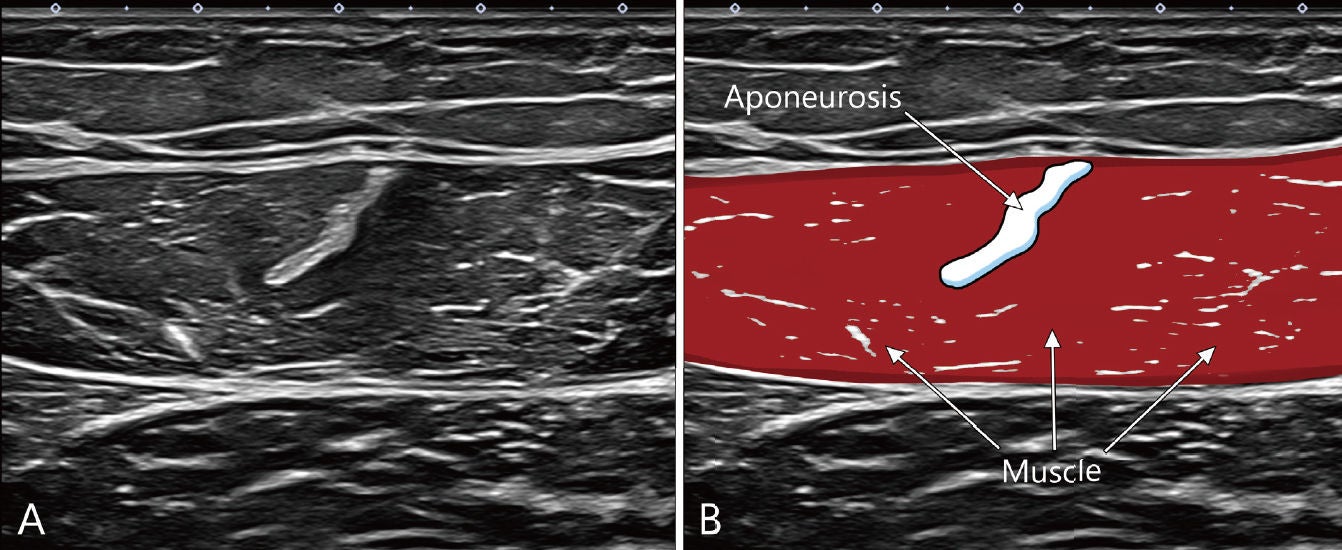 Fig. 3: Short-axis ultrasound image of the rectus femoris and its intramuscular aponeurosis. On ultrasound, muscle fi bers display a starry-sky pattern: a hypoechoic background corresponding to the muscle fi bers, with hyperechoic dots and lines corresponding to the perimysium. The aponeurosis (connective tissue) appears hyperechoic with a highly homogeneous salt-and-pepper pattern and well-defi ned borders.
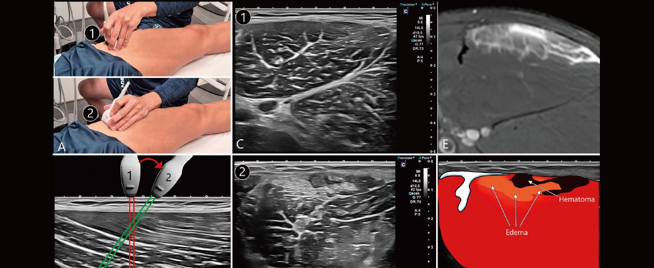 Fig. 4: Muscle edema in a rectus femoris injury. (A) Diff erent probe orientations. (B) Long-axis ultrasound image of the rectus femoris showing fi ber direction and the angle of the ultrasound beam relative to the fi bers. (C) Ultrasound image of the rectus femoris with the probe in position 1, perpendicular to the long axis of the thigh; the connective tissue is well defi ned, but muscle edema is not visible. (D) Same case, same examination, with the probe in position 2 (angled cranially); the edema is clearly visible, with a superfi cial hematoma. (E) Proton density fat-saturated (PD-FS) sequence obtained the same day, showing a distribution similar to that seen on ultrasound. (F) Same image as in D, with relevant structures highlighted.
Connective tissue injuries
In recent years, the involvement of muscle connective tissue has gained increasing relevance, to the point that a direct relationship is now recognized between the extent and location of affected connective tissue and the prognosis of muscle injury.9, 10, 11
The work of Balius, Pedret and co-workers on the histology of connective tissue and its involvement in muscle injury has described important differences between tendon, aponeurosis and fascia.9, 10
Understanding this organization is essential to the assessment and management of muscle injury.
Tendons connect muscle to bone. They are composed of dense regular connective tissue in which all collagen fibers are aligned in the same direction, which allows them to withstand high longitudinal tensile forces.10
Aponeuroses are also composed of dense connective tissue, similar to that of tendon, but with different characteristics. Their collagen fibers are organized in distinct layers with varying orientations between them, allowing for force dispersion and enabling the structure to withstand both longitudinal and transverse tensile loads. Some aponeuroses are located at the periphery of the muscle while others are intramuscular.
Fasciae surround the muscles and differ structurally from both aponeuroses and tendons. They are composed of loose connective tissue with greater flexibility.10
This distinction has practical implications. Tendon involvement is associated with a longer recovery time than aponeurotic injury, and injuries involving the myotendinous junction carry a higher risk of re-injury.10,16
Myofascial junction injuries and purely intramuscular injuries generally carry a better prognosis and a lower risk of re-injury.
Each muscle has a distinct distribution of connective tissue, and injuries tend to occur at locations characteristic of each muscle. Knowing these patterns before scanning, and examining the most vulnerable areas in greater detail, significantly improves the diagnostic yield of the examination.11, 12, 13
Several classification systems have been developed to translate these anatomical findings into reproducible prognostic categories. Nomenclature based on the histoarchitecture of the connective tissue sheath provides the common framework,9, 10 while muscle-specific ultrasound classifications have been proposed for the medial gastrocnemius,11 the biceps femoris T-junction,12 the proximal hamstring origin tendon,14, 15 and the quadriceps.17 MRI-based classification of muscle healing complements these systems during the later stages of follow-up.18 Familiarity with the system applicable to each muscle allows sonographic findings to be reported in terms that carry direct prognostic meaning.
Assessment of possible connective tissue involvement should be performed separately from the assessment of muscle tissue. Frequency, depth, focus, and gain must be readjusted to achieve the best possible image quality.
The axial plane is primarily used, with the probe oriented perpendicular to the direction of the tendon or aponeurosis being examined.
Tendons and aponeuroses appear as hyperechoic structures with marked anisotropy, displaying a very uniform internal pattern with well-defined margins, known as a “salt-and-pepper” pattern (Fig. 3). Very slow and meticulous sweeps should be performed, seeking areas of differing echogenicity within the structure, with small probe angulations in suspicious areas to assess their anisotropic behavior.
When a tendon or aponeurosis is injured, the homogeneity of its pattern is disrupted, with internal hypoechoic or isoechoic areas that may be associated with thickening and loss of anisotropy (Figs. 5, 6). These findings are accompanied by edema in the adjacent muscle fibers, which may be subtle or, in some cases, hemorrhagic.
In superficial locations, the use of higher-frequency probes may be recommended to achieve greater resolution (Fig. 5).
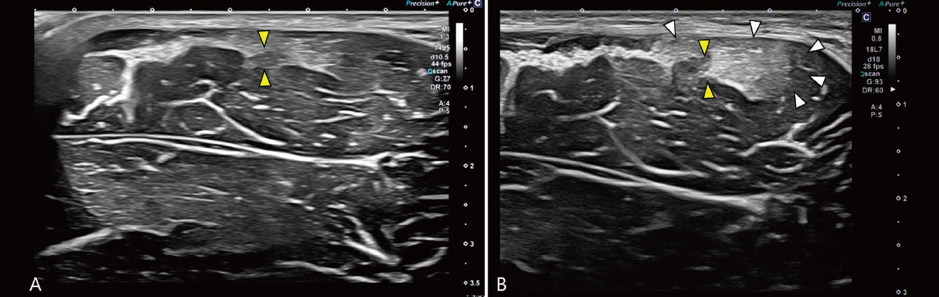 Fig. 5: Injury of the intramuscular tendon of the adductor longus: same-day comparative ultrasound with different probes. (A) PLU-1005BT probe. (B) PLU-1204BT probe. Better delineation of tendinous involvement (yellow arrowheads) and edema (white arrowheads) is observed with the higherfrequency probe.
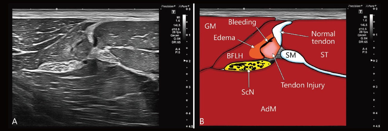 Fig. 6: Injury of the conjoint tendon of the biceps femoris and semitendinosus. (A) Ultrasound image. (B) Same image with relevant structures highlighted. The difference between the healthy and the injured tendon is clearly appreciated. GM, gluteus maximus; BFLH, biceps femoris long head; ST, semitendinosus; ScN, sciatic nerve; AdM, adductor magnus.
In severe injuries with clear tendon discontinuity, the disruption is typically seen on short-axis imaging as an abrupt hypoechoic thickening of the tendon (the tendon stump) immediately before it disappears (Fig. 6). As an indirect sign of rupture, acoustic shadowing may occur due to critical-angle artifacts, both at the tendon stump itself and at areas of tendon undulation caused by loss of tension and retraction. These are more clearly visible in the longitudinal plane (Fig. 7). Care must be taken not to confuse these critical-angle shadows with calcifications that may develop during the healing of some tendinous injuries or with myositis ossificans. In these cases, the shadows are denser and arise from a hyperechoic interface at their upper margin (Fig. 8).
Injuries located in deep regions or within old scar tissue, particularly when small, are difficult to interpret even for the most experienced sonographers.7
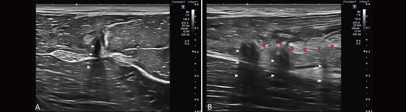 Fig. 7: Tendon injury with discontinuity and a critical-angle shadowing artifact. (A) Axial ultrasound image. (B) Longitudinal ultrasound image. Acoustic shadows caused by loss of tendon tension are visible (white arrowheads); on the longitudinal view, tendon waviness can also be appreciated (red arrowheads).
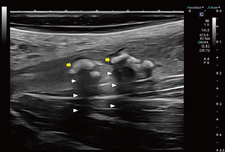 Fig. 8: Calcifi cations during healing of an adductor longus tendon injury. Acoustic shadowing (white arrowheads) and the hyperechoic edge of the calcifi cation (yellow arrows) are seen.
Presence of hemorrhage
Acute muscle injuries are frequently accompanied by hemorrhage, which can vary widely in volume depending on the structures involved. Hemorrhage typically spreads toward zones of least resistance, such as fascial or interaponeurotic planes. Some injuries have a marked tendency toward bleeding and hematoma formation, such as myoaponeurotic injuries of the medial gastrocnemius or avulsions of the adductors or hamstrings, while others bleed considerably less, either due to their volume or location, such as injuries to the conjoint tendon of the biceps femoris and semitendinosus or lesions in the central septum of the rectus femoris. Hematomas are most easily detected 48–72 hours after injury, when liquefaction of their contents causes them to appear as anechoic collections with generally well-defi ned walls, compressible under the probe (Fig. 9). In some cases the hematoma appears clotted, producing a diff erent appearance, with areas of intermediate echogenicity corresponding to clot against an anechoic background of unclotted blood (Fig. 10).
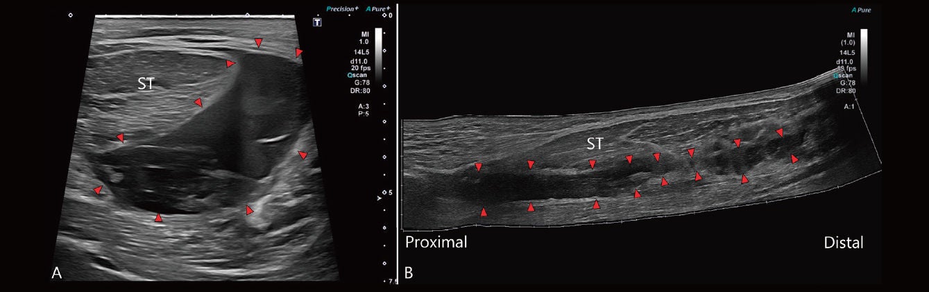 Fig. 9: Liquefi ed hematoma in a semimembranosus avulsion. (A) Axial view. (B) Panoramic longitudinal reconstruction. A hypo- to anechoic area corresponding to non-clotted blood is observed (red arrowheads). ST, semitendinosus.
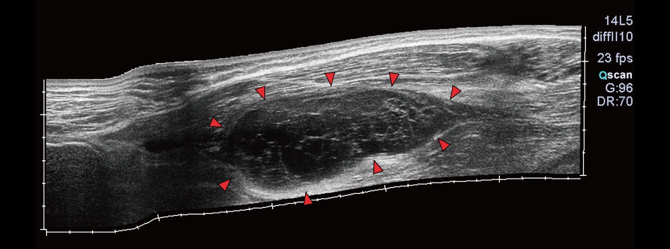 Fig. 10: Panoramic reconstruction of a clotted hematoma from a semimembranosus avulsion (red arrowheads). Note the isoechoic areas within the hematoma, indicating clot formation and therefore precluding evacuation by ultrasound-guided aspiration.
Intramuscular hematomas are generally less voluminous than interaponeurotic ones or avulsion-related hematomas and appear as hypoechoic (acute) or anechoic (subacute) collections with more irregular margins (Fig. 4D). Interfascial hemorrhage has a distinct sonographic appearance. It typically presents as hyperechoic or isoechoic thickening of the fascial plane between two muscles, or between muscle and subcutaneous tissue (Fig. 11).12 These are more diffi cult to detect, especially in deep planes, yet they provide highly valuable information, as they appear very early and are evidence of a structural injury that may be located at a distance and must be identifi ed.
Although hematomas tend to resorb over the medium to long term, in our practice voluminous collections are drained under ultrasound guidance with the aim of promoting better healing.20 Once the hematoma has clotted, needle aspiration is no longer feasible and alternative strategies must be considered.
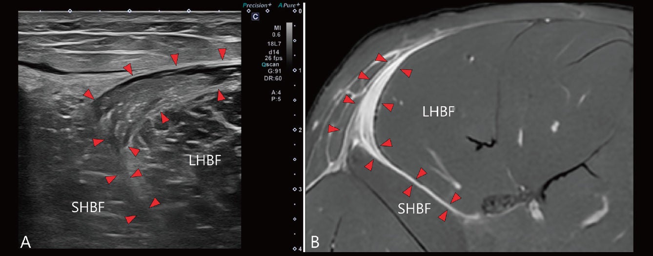 Fig. 11: Interfascial bleeding. (A) Axial ultrasound image obtained 48 h after an injury at the T-junction between the long and short heads of the right biceps femoris. (B) Axial PD-FS magnetic resonance image obtained at 72 h. Isoechoic thickening of the upper border of the biceps femoris long head, extending between the long and short heads, is appreciated (red arrowheads).
Use of ultrasound for muscle injury assessment in a professional soccer team
The use of ultrasound for the assessment of muscle injuries in a professional soccer team should cover the following aspects: initial evaluation following injury, monitoring throughout the rehabilitation process with early detection of complications, and, finally, guidance for interventional procedures.
Initial assessment of a muscle injury with ultrasound
As noted above, decision-making in professional soccer is subject to considerable time pressure. The player and the coaching staff need to know the severity of the injury as soon as possible, and waiting 24–48 hours for initial information is rarely acceptable.
At Athletic Club, we perform ultrasound as early as possible—depending on the timing of the injury (match or training session), sometimes within just a few minutes (Fig. 12), and in other cases the following day—but it is rare for the ultrasound examination to be delayed beyond 24 hours, thereby achieving an early diagnosis with highly relevant information in the majority of cases.
The examination is repeated the following day, since, as previously discussed, edema and hemorrhage are more conspicuous at 24–48 hours.
To obtain the best results, we always incorporate ultrasound within the overall clinical assessment of the player as an integral part of the diagnostic process.
To this end, a clinical history is taken to identify the type of trauma (direct or indirect), the mechanism of injury (stretching, sprinting, kicking, or other), the onset pattern (acute explosive onset with a clearly identifiable trigger, or progressive overload without an identifiable cause), and the location as reported by the player.
We place particular importance on pain intensity, which in our experience correlates with prognosis, although it has not been validated as an independent predictor. Pain reported by the player on days 1, 2 and 3 after injury is systematically recorded using a visual analog scale (VAS).
Following the history, a working diagnostic hypothesis is established, which is then refined through a clinical examination aimed at more precisely localizing the potentially involved structures using provocative tests (resisted testing, stretching) and direct palpation of painful areas.
Combining clinical history, physical examination, current knowledge of the different types of muscle injury and a meticulous ultrasound technique, with particular attention to the highest-risk areas, has proved reliable in our practice. However, this focused clinical approach can occasionally lead to error by directing the examination too narrowly and overlooking other areas. For this reason, in addition to the above, a general systematic approach should also be followed to allow for a more comprehensive assessment of all potentially involved structures, thereby reducing the likelihood of false negatives.
Finally, in moderate or severe injuries, high-field MRI (1.5 T or 3 T) is also obtained to achieve greater definition and accuracy regarding the structures involved.18
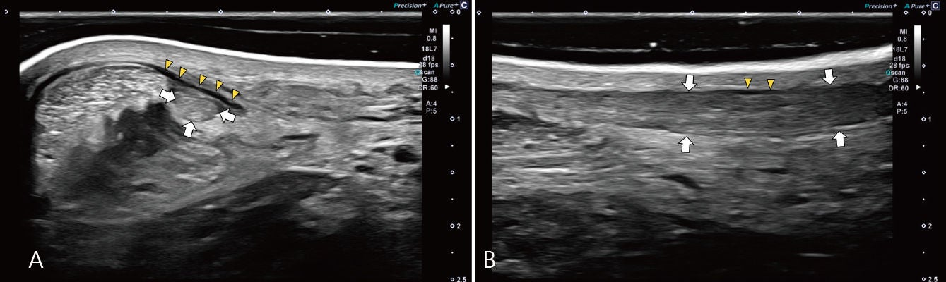 Fig. 12: Ultrasound performed a few minutes after an injury sustained during training. The player reported acute pain and functional impairment. (A) Axialview. (B) Longitudinal view. Thickening of the plantaris tendon is seen (white arrows), surrounded by a thin layer of fl uid (yellow arrowheads). MRI was not considered necessary in this case.
Monitoring muscle injuries
We consider one of the primary functions of ultrasound in muscle injury to be longitudinal follow-up. For this purpose, a systematic weekly ultrasound examination is performed until return-to-play clearance in mild and moderate injuries. In severe injuries, an MRI follow-up is additionally obtained at week 4.18
During recovery from a muscle injury, the player frequently experiences discomfort that may either be normal or indicative of a problem. In other cases, the injury may evolve unfavorably without the player reporting any adverse symptoms. Performing an MRI every time there is some discomfort, or on a repetitive basis, is not practical; however, ultrasound is ideally suited to fulfill this function.
The maturational changes of scar tissue are not adequately assessed by ultrasound, which cannot reliably distinguish between the phases of scar maturation at 4, 6 or 8 weeks. MRI is clearly superior for this purpose.18
The role of ultrasound in the follow-up of muscle injury is therefore not to assess the maturity of scar tissue, but to detect complications early.
Persistent muscle edema, increasing or new hemorrhage, and the appearance of myositis ossificans are warning signs that the tissues are not adapting to the imposed loads and that the risk of re-injury is high (Fig. 13).19 In these cases we slow the progression of rehabilitation, intensify follow-up, and perform MRI when the suspicion of re-injury is high.
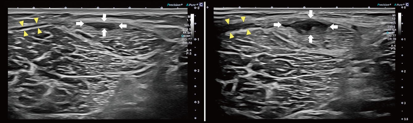 Fig. 13: Follow-up of muscle injuries. Comparative axial ultrasound images of the biceps femoris short head obtained one week apart. The player reported discomfort following an involuntary overstretch during the late rehabilitation phase. The ultrasound shows an increase in the area of injury (white arrows) and in fascial bleeding (yellow arrowheads).
Use of ultrasound in interventional procedures
Ultrasound is an extremely useful tool for evaluating hematomas, as well as for guiding their drainage to promote better healing (Fig. 14). In selected cases it may be used to guide the injection of growth factors, for example in severe injuries or those following an unfavorable course.20 The use of ultrasound ensures accurate placement of the product at the precise site of injury and enhances procedural safety by enabling avoidance of critical adjacent structures such as vessels and nerves.
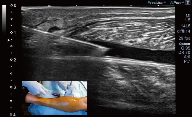 Fig. 14: Ultrasound-guided intervention: evacuation of a hematoma between the gastrocnemius and the soleus.
Limitations
Ultrasound assessment of muscle injury has well-recognized limitations. It is highly operator-dependent, with reported interobserver variability that is greater for small, deep or intratendinous lesions.
Examinations performed very early after injury may yield false negatives, since edema and hemorrhage become more conspicuous at 24–48 hours; a normal early scan therefore does not exclude a structural injury and should be repeated.
Ultrasound cannot reliably characterize scar maturation, and it does not replace MRI in severe injuries or when surgical management is being considered.
Finally, the fi ndings and protocols described here refl ect the experience of a single institution and have not been subjected to formal diagnostic accuracy analysis against MRI.7, 16
Conclusions
Ultrasound is a highly useful technique in muscle injuries when applied correctly.
Optimal results require an experienced musculoskeletal sonographer, a high-quality ultrasound system with probes suited to each type of injury, integration of ultrasound findings within the clinical context of the athlete, and use of MRI whenever necessary.
Ultrasound enables serial examinations to be performed in a comfortable and efficient manner, allowing early detection of adverse clinical evolution, exclusion of recurrent injury, and guidance of interventional procedures.
All these applications have made ultrasound an indispensable tool in a professional soccer team.
Conflict of interest
This white paper was produced with the support of Canon Medical Systems Europe B.V.
The ultrasound systems and transducers described are Canon products. The author has received an honorarium from Canon Inc. for the preparation of this paper.
Canon Inc. and Canon Medical Systems Europe B.V. had no role in the selection of clinical cases, the interpretation of imaging findings, or the conclusions expressed. The clinical material and protocols described correspond to the author’s practice within the Medical Department of Athletic Club, Bilbao, and have not been subjected to independent peer review.
References
- Ekstrand J, Hägglund M, Waldén M.
Epidemiology of muscle injuries in professional football (soccer). Am J Sports Med. 2011;39(6):1226–32.
doi:10.1177/0363546510395879 · PMID 21335353
- Ekstrand J, Spreco A, Bengtsson H, Bahr R.
Injury rates decreased in men’s professional football: an 18-year prospective cohort study of almost 12 000 injuries sustained during 1.8 million hours of play. Br J Sports Med. 2021;55(19):1084–91.
doi:10.1136/bjsports-2020-103159 · PMID 33547038
- Bahr R, Clarsen B, Ekstrand J.
Why we should focus on the burden of injuries and illnesses, not just their incidence. Br J Sports Med. 2018;52(16):1018–21.
doi:10.1136/bjsports-2017-098160 PMID 29021247
- Ekstrand J, Bengtsson H, Waldén M, Davison M, Khan KM, Hägglund M.
Hamstring injury rates have increased during recent seasons and now constitute 24% of all injuries in men’s professional football: the UEFA Elite Club Injury Study from 2001/02 to 2021/22. Br J Sports Med. 2023;57(5):292–8.
doi:10.1136/bjsports-2021-105407 · PMID 36588400
- Palermi S, Scavone A, Anzà M, Gregorace E, Vecchiato M, Bordalo M, Forster BB, Jacobson JA, Myers C, Iriarte I, Pedret C.
Diagnostic ultrasound of muscle injuries: what the sports medicine clinician should know. Br J Sports Med. 2025;59(24).
doi:10.1136/bjsports-2025-110255 · PMID 40967835
- Zunarelli P, Lucenteforte G, Miceli M, Stride M, Della Villa F.
The use of diagnostic ultrasound in sports muscle injuries in football (soccer) players: state-of-the-art review. Curr Phys Med Rehabil Rep. 2022;10:154–62.
doi:10.1007/s40141-022-00354-7
- Paoletta M, Moretti A, Liguori S, Snichelotto F, Menditto I, Toro G, Gimigliano F, Iolascon G.
Ultrasound imaging in sport-related muscle injuries: pitfalls and opportunities. Medicina (Kaunas). 2021;57(10):1040.
doi:10.3390/medicina57101040 · PMID 34684077
- Iriarte I, Pedret C, Balius R, Cerezal L.
Ultrasound of the musculoskeletal system: anatomical exploration and pathology. Bilbao: Mskroom; 2021.
Available from: https://mskroom.com/
- Balius R, Blasi M, Pedret C, Alomar X, Peña-Amaro J, Vega JA, Pruna R, Ardèvol J, Álvarez G, de la Fuente J, Fernández-Jaén T, Järvinen TAH, Rodas G;
Study Group of the Muscle and Tendon System, Spanish Society of Sports Traumatology. A histoarchitectural approach to skeletal muscle injury: searching for a common nomenclature. Orthop J Sports Med. 2020;8(3):2325967120909090.
doi:10.1177/2325967120909090 · PMID 32232071
- Pedret C, Peña-Amaro J, Balius R, Järvinen T.
Histological definition of skeletal muscle injury: a guide to nomenclature along the connective tissue sheath/structure. Sports Med. 2025;55(5):1061–5.
doi:10.1007/s40279-024-02165-3 · PMID 39692975
- Pedret C, Balius R, Blasi M, Dávila F, Aramendi JF, Masci L, de la Fuente J.
Ultrasound classification of medial gastrocnemius injuries. Scand J Med Sci Sports. 2020;30(12):2456–65.
doi:10.1111/sms.13812 · PMID 32854168
- Cronin K, Kerin F.
Ultrasound-based classification and rehabilitation of biceps femoris T-junction injuries. Front Sports Act Living. 2026;8:1735177.
doi:10.3389/fspor.2026.1735177 · PMID 41710092
- Becciolini M, Bonacchi G, Bianchi S.
Ultrasound features of the proximal hamstring muscletendon- bone unit. J Ultrasound Med. 2019;38(6):1367– 82.
doi:10.1002/jum.14804 · PMID 30260012
- Wada M, Kumai T, Okunuki T, Sugimoto T, Ishizuka K, Tanaka Y.
Ultrasound diagnosis of hamstring muscle complex injuries focused on origin tendon structure: male university rugby players. Diagnostics (Basel). 2025;15(1):54.
doi:10.3390/diagnostics15010054 · PMID 39795581
- Wada M, Okunuki T, Sugimoto T, Tanaka Y, Kumai T.
Impact of proximal conjoint tendon injury on return to play in the BF-ST complex: a prospective MRI-based study. Diagnostics (Basel). 2026;16(1):166.
doi:10.3390/diagnostics16010166 · PMID 41515661
- Petersen J, Thorborg K, Nielsen MB, Skjødt T, Bolvig L, Bang N, Hölmich P.
The diagnostic and prognostic value of ultrasonography in soccer players with acute hamstring injuries. Am J Sports Med. 2014;42(2):399–404.
doi:10.1177/0363546513512779 · PMID 24335495
- Manske RC, Voight M, Wolfe C, Page P.
The use of MSK ultrasound with quadriceps muscle injury. Int J Sports Phys Ther. 2023;18(1):282–4.
doi:10.26603/001c.68077 · PMID 36798902
- Isern-Kebschull J, Mechó S, Pedret C, Pruna R, Alomar X, Kassarjian A, Luna A, Martínez J, Tomas X, Rodas G.
Muscle healing in sports injuries: MRI findings and proposed classification based on a single institutional experience and clinical observation. Radiographics. 2024;44(8):e230147.
doi:10.1148/rg.230147 · PMID 39052498
- Drakonaki EE, Sudoł-Szopińska I, Sinopidis C, Givissis P.
High resolution ultrasound for imaging complications of muscle injury: is there an additional role for elastography? J Ultrason. 2019;19(77):137–44.
doi:10.15557/JoU.2019.0020 · PMID 31355586
- Martínez-Zapata MJ, Orozco L, Balius R, Soler R, Bosch A, Rodas G, Til L, Peirau X, Urrútia G, Gich I, Bonfill X; PRP-RICE Group.
Efficacy of autologous platelet-rich plasma for the treatment of muscle rupture with haematoma: a multicentre, randomised, double-blind, placebo-controlled clinical trial. Blood Transfus. 2016;14(2):245–54.
doi:10.2450/2015.0099-15 · PMID 26509827
Social Media


