

Ultrasound / White paper
Ultrasound 3rd Harmonic Imaging: Challenges and benefits

Introduction
Ultrasound is a highly useful imaging modality in both clinical and research settings. For this reason, it is essential to deepen our understanding and improve the tools that enhance image resolution. Although ultrasound is sometimes considered controversial due to its operator dependency, advancements in data processing and artificial intelligence have significantly improved image resolution and diagnostic accuracy. With better tools becoming available, diagnoses are increasingly objective and reliable.
The single-crystal PVI-475BX transducer, equipped with intelligent Dynamic Micro-Slice (iDMS) technology, offers improved image quality and aids in diagnosis without extending examination time. In this paper, we present the physical principles that help explain the concepts behind third harmonic image acquisition and provide clinical cases where its usefulness has been demonstrated—such as in lesion characterization, identifying acute inflammation, and more. We hope this information proves valuable and contributes to enhancing diagnostic performance in ultrasound practice.
Canon’s innovative technology“Third Harmonic Imaging”
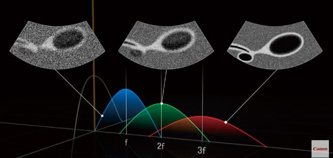
Progress of harmonic technology by Canon
When second harmonic frequencies (generated by frequency doubling) are received and imaged, only the central portion of the ultrasound beam—where harmonics are most likely to occur—is utilized. This results in high resolution images with fewer artifacts. This technique, known as Tissue Harmonic Imaging (THI), is used across various clinical applications and is incorporated in many ultrasound systems. It is well known that not only frequency doubling (second harmonic) components but also triple frequency (third harmonic) components are reflected by the body. It was hypothesized that leveraging third harmonics could enhance the benefits of THI. However, due to the lower signal strength and the interference from noise, third harmonic signals have been difficult to isolate. Canon developed an advanced method to extract these weak third harmonic signals by integrating its iBeam+ beamforming technology with AI-enabled design*. This innovation led to the successful commercialization of the world’s first ultrasound imaging method using third harmonic waves, known as 3rd Harmonic Imaging (3-HI).
* AI technology is used at the design stage. The system does not have a self-learning function.
Clinical Cases
Case 1
A 33-year-old asymptomatic woman undergoing follow-up for gallbladder polyps. On the conventional ultrasound image, two broad-based polyps are seen attached to the anterior gallbladder wall, with the largest measuring 6 mm (Fig. 1a). In the 3-HI image, three anterior and two posterior wall polyps are identified. Broad-based in the wall and sessile morphology are clearly defined (Fig.1b). No focal wall thickening is observed. These polyps are considered low-risk for malignancy and, given their size (<9 mm), do not require follow-up.
Accurate evaluation of gallbladder polyps includes assessing their morphology, size, and relationship with the gallbladder wall (broad or narrow base). These features are better defined with third harmonic imaging, which also enables more reliable assessment of wall thickness—an important criterion for categorizing polyps, as per the Ultrasound Radiologists Consensus.
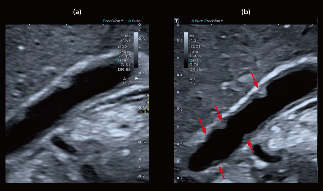
Figure 1 (a) Conventional image
(b) 3-HI image
Case 2
A 23-year-old woman presented to the emergency department with right upper quadrant abdominal pain. The conventional ultrasound showed thickening of the anterior gallbladder wall and biliary sludge (Fig. 2a).
However, the 3-HI image revealed a thin-walled gallbladder with biliary sludge, helping to rule out gallbladder inflammation as the cause of pain (Fig. 2b).
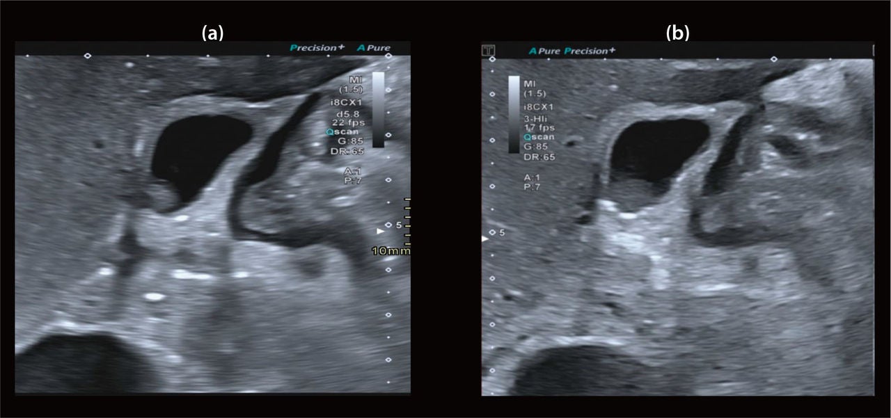
Figure 2 (a) Conventional image
(b) 3-HI image
Case 3
A 36-week pregnant woman with abdominal pain and a history of cholelithiasis. The conventional image showed cholelithiasis and apparent gallbladder wall thickening (Fig.3a). In contrast, the 3-HI image clearly defined the gallbladder wall and confirmed the presence of cholelithiasis without signs of acute cholecystitis (Fig.3b).
The improved spatial resolution and reduced speckle noise in the 3-HI image allowed better delineation of wall contours, crucial in diagnosing acute cholecystitis.
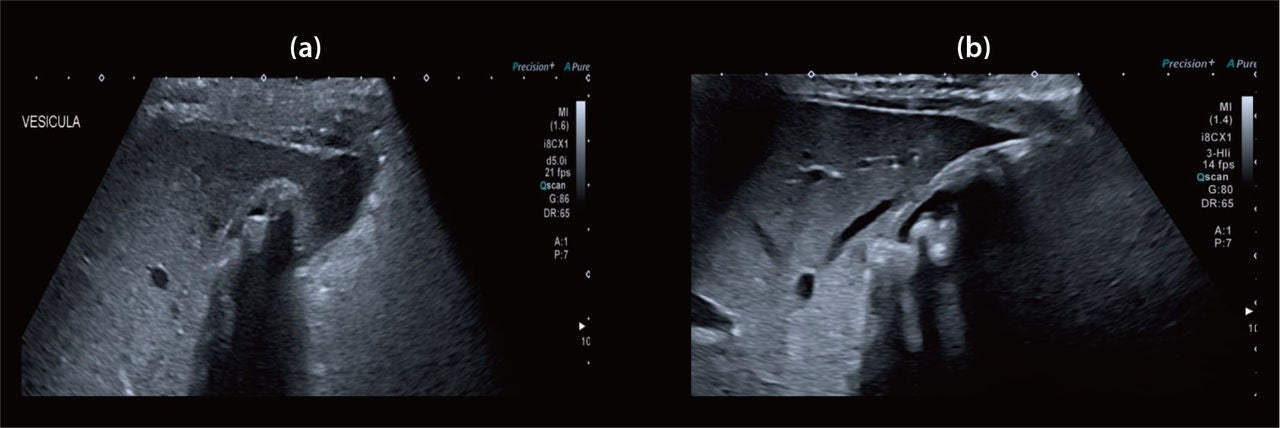
Figure 3 (a) Conventional image
(b) 3-HI image
Case 4
A 70-year-old with no medical history, presented with gastrointestinal bleeding. The conventional ultrasound showed a liver with heterogeneous echotexture and a normal anterior capsule (Fig.4a). The 3-HI image revealed the same parenchymal heterogeneity but also showed anterior surface lobulation suggestive of advanced chronic liver disease (Fig.4b).
This highlights the benefit of enhanced axial and lateral resolution in 3-HI images, allowing better definition of capsular lobulation as depicted also in case 5.
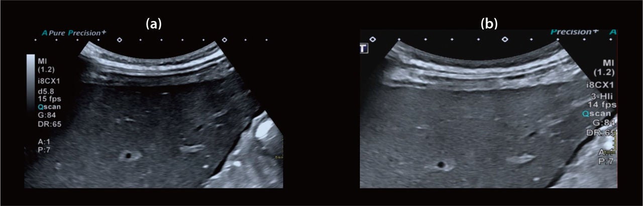
Figure 4 (a) Conventional image
(b) 3-HI image
Case 5
A 55-year-old asymptomatic woman with a history of breast cancer and abnormal liver function tests. Conventional ultrasound revealed heterogeneous echogenicity with limited posterior visualization (Fig.5a). The 3-HI image provided better depth resolution, showing a smooth anterior capsule and an undulating posterior capsule up to 6 mm, suggestive of chronic liver disease (Fig.5b). The diagnosis of primary biliary cholangitis was later confirmed. This technique was also valuable in pediatric liver parenchyma assessment.
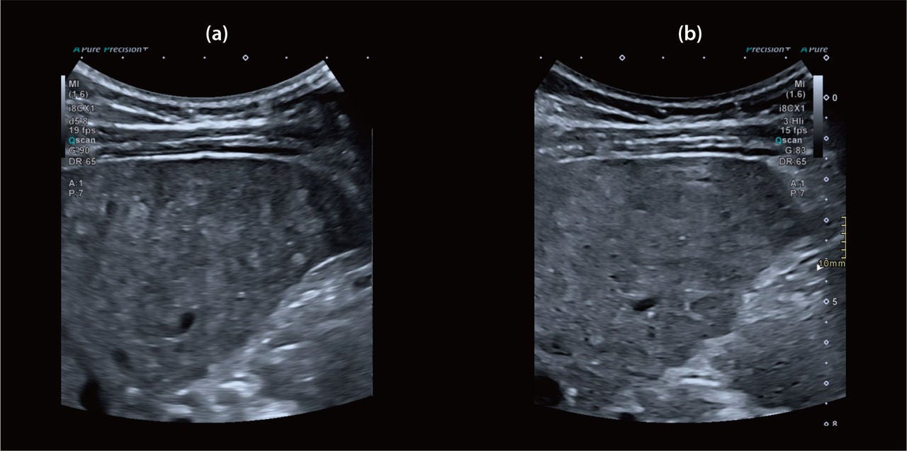
Figure 5 (a) Conventional image
(b) 3-HI image
Case 6
A 6-year-old girl with a liver abscess due to Ascaris-induced bile duct obstruction.
Conventional imaging showed a subcapsular abscess in liver segment VII ( ↑ ) (Fig. 6a). The 3-HI image enhanced the visibility of the abscess ( ↑ ) and increased echogenicity of intrahepatic bile ducts ( ↓ ) consistent with acute cholangitis (Fig. 6b).
Zoomed-in conventional image shows heterogeneous liver parenchyma and the abscess contacting the right hepatic vein (Fig. 6c). Zoomed-in 3-HI image further confirmed these findings, and showed clearer abscess boundaries ( ↑ ) and bile duct structures ( ↑ ) (Fig. 6d).
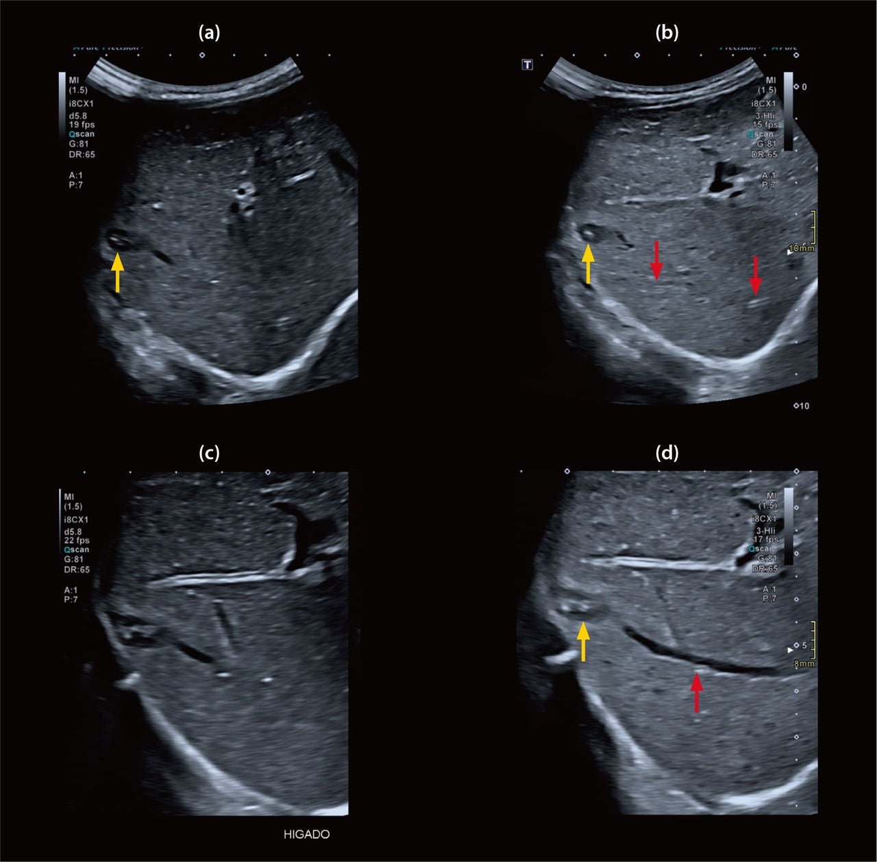
Figure 6 (a) Conventional image
(b) 3-HI image
(c) Conventional image zoom
(d) 3-HI image zoom
Case 7
A 42-year-old asymptomatic woman under follow-up for hepatic steatosis and a focal lesion. Conventional ultrasound showed diffuse increased parenchyma echogenicity, anterior capsule lobulation and a hypoechoic lesion between liver segments IVB and VIII (Fig. 7 a). The 3-HI image better depicted steatosis and capsule changes (diffuse parenchyma increased echogenicity was higher and better depicted) (Fig. 7b). MRI confirmed the lesion as focal nodular hyperplasia.
T2-weighted fat saturation MRI anterior capsule lobulation due to a focal lesion between liver segments IVB and VIII, with a slightly hyperintense peripheral component and a hyperintense central scar, a finding consistent with a focal nodular hyperplasia (Fig. 7c). T1-weighted post-contrast late-phase MRI shows anterior capsule lobulation due to a nodular lesion with late enhancement of the central scar (Fig. 7d).
*Increased comparative echogenicity is also found in 3-HI images when heterogeneous fluid content is evaluated, as seen in the following case.
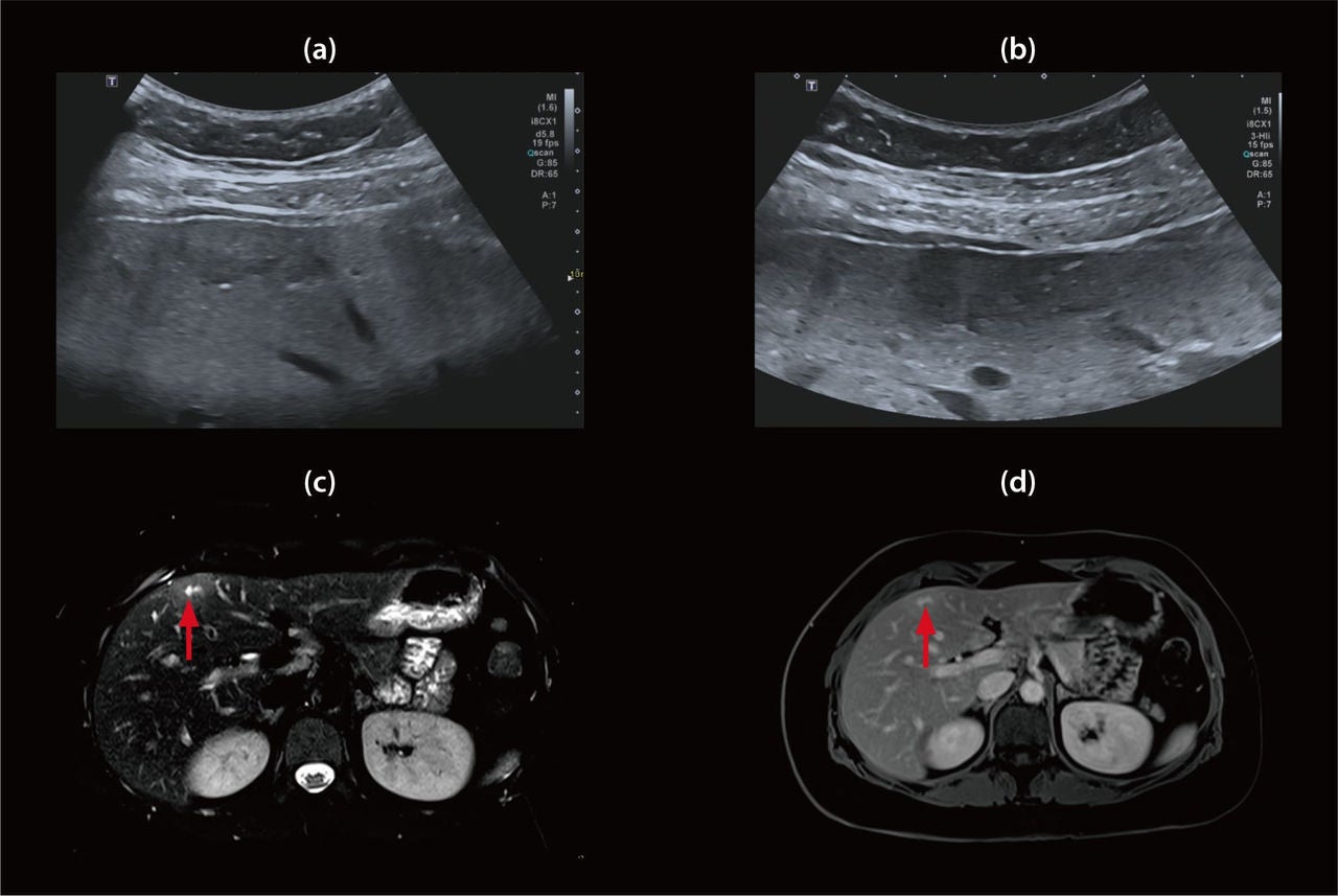
Figure 7 (a) Conventional image
(b) 3-HI image
(c) T2-weighted fat saturation MRI
(d) T1-weighted post contrast late-phase MRI
Case 8
A 70-year-old man presented with urinary obstruction due to benign prostatic hyperplasia. Urinary infection and abundant urine sediment were found. Conventional ultrasound showed bladder wall thickening, prostate enlargement, and echogenic particulate content (Fig. 8a). The 3-HI image provided a clearer view of these features and revealed echogenic bands between the abdominal wall and bladder, likely due to tissue heterogeneity.
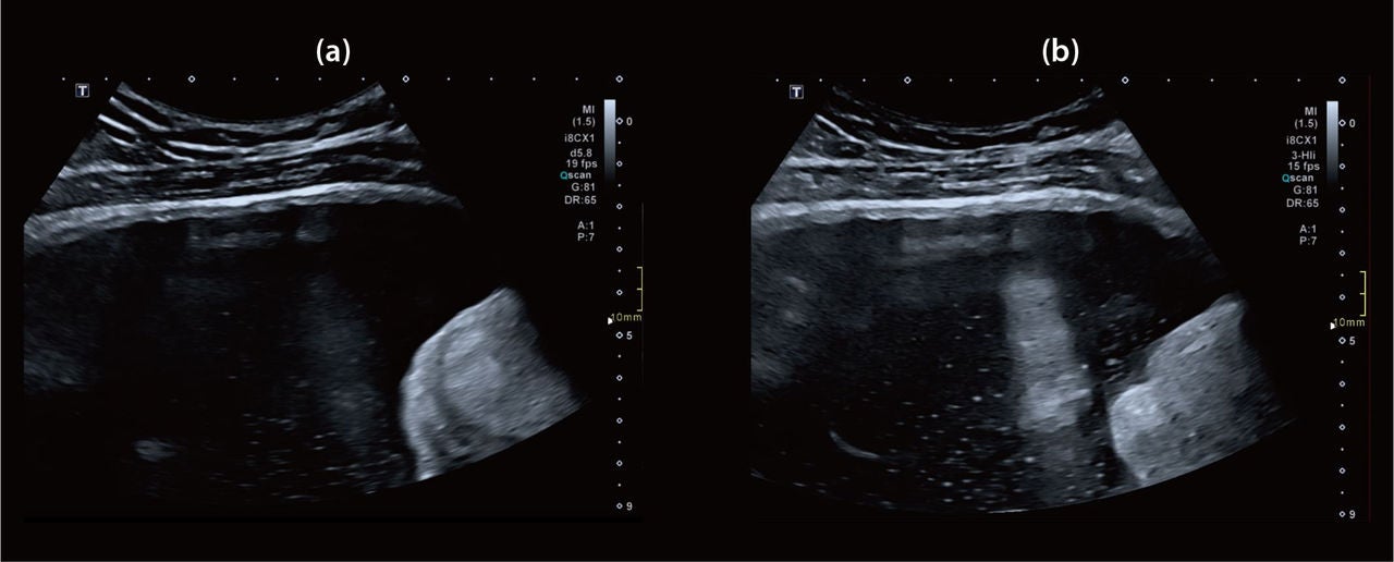
Figure 8 (a) Conventional image
(b) 3-HI image
Challenges in interpreting 3-HI images
In the presented cases, increased echogenicity of the liver parenchyma was observed with 3-HI imaging. This represents one of the main challenges in interpretation and underscores the importance of accurately identifying and characterizing hepatic steatosis (as in Case 7) and acute inflammatory processes.
This finding may be explained by the following factors: Improved penetration, resulting in greater contrast between the parenchyma and adjacent tissues, as observed in Case 6. Artifact reduction, which makes the parenchyma appear more homogeneous and uniform.
Due to its heterogeneous nature, liver parenchyma can generate a large number of harmonics, contributing to the increased echogenicity seen in Case 7.
Conclusion
The iDMS matrix probe with 3-HI technology provides superior image quality and resolution, providing more information particularly when evaluating the gallbladder and liver parenchyma. However, optimizing its use requires understanding of ultrasound physics and recognizing possible artifacts introduced by high-resolution imaging and tissue composition. AI-enabled tools, such as 3-HI, and data post processing improve diagnostic performance without increasing scan time. Ongoing experience sharing and data collection will further enhance the clinical value of this technology.
Recommended readings
- Amer M, Sharma R, Knipe H. SRU management guidelines for gallbladder polyps. Reference article, Radiopaedia.org (Accessed on 17 Mar 2025) https://radiopaedia.org/articles/186677.
- Sundaram KM, Morgan MA, Depetris J, et al. Imaging of benign gallbladder and biliary pathologies in pregnancy. Abdom Radiol 48, 1921–1932 (2023). https://doi.org/10.1007/s00261-023-03832-1
- Zheng T, Qu Y, Chen J, et al. Noninvasive diagnosis of liver cirrhosis: qualitative and quantitative imaging biomarkers. Abdom Radiol 49, 2098–2115 (2024). https://doi.org/10.1007/s00261-024-04225-8
- Di Serafino M, Gioioso M, Severino R, et al. Ultrasound findings in paediatric cholestasis: how to image the patient and what to look for. J Ultrasound 23, 1–12 (2020). https://doi.org/10.1007/s40477-019-00362-9
- Fouladi D, Shao N, Zarghampour M, Pandey A, Pandey P, Kamel IR (2018). Radiographic Assessment for Liver Tumors. In: Cardona, K., Maithel, S. (eds) Primary and Metastatic Liver Tumors. Springer, Cham. https://doi.org/10.1007/978-3-319-91977-5_2
- Radiology 2022; 305: 277-289. Management of incidentally detected Gallbladder Polyps: Society of Radiologists in Ultrasound Consensus conference Recommendations.
Social Media


