

Ultrasound / White paper
User experience of the Aplio i-series / Prism Edition for pancreatobiliary disease

Introduction
High-end ultrasound systems can provide value in the pancreatobiliary environment. Ultrasound accessibility and availability for patients is a strength; in addition, the latest ultrasound systems can see more information. Canon’s Aplio i-series / Prism Edition provides new technology solutions for pancreatobiliary assessment. The system is built around the core technology, iBeam+, an advanced beamforming technology that can perform high-speed processing for huge quantities of received data. This inherent technology has led to developments with real value in pancreatobiliary imaging.
The pancreas has always been somewhat of a challenge for ultrasound. The Aplio i-series / Prism Edition provides excellent B-mode resolution, which is a big strength; however, the introduction of a new harmonic technique, 3rd Harmonic Imaging (3-HI), which builds on the core iBeam+ technology provides even more detail. A dedicated convex interventional transducer, combined with existing tools like Superb Micro-vascular Imaging (SMI), provides significant clinical value to the pancreatobiliary clinician.
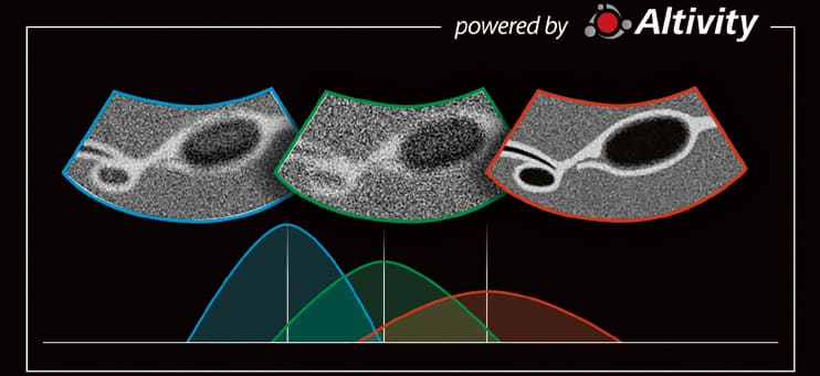
Figure 1 Progress of harmonic technology
*AI technology is used at the design stage. The system does not have a self-learning function.
Efficiently extracting the 3rd harmonic components in ultrasound represented a complex challenge that Canon overcame by utilizing AI during the design stage of the system. An AI filter for 3rd harmonics was developed using deep learning that effectively allows the extraction and utilization of the 3rd harmonic signal, which had conventionally been difficult to achieve. The 3-HI can provide higher resolution, contrast, and less noise than the traditional 2nd harmonic imaging, resulting in sharper, clearer images (Fig. 1).
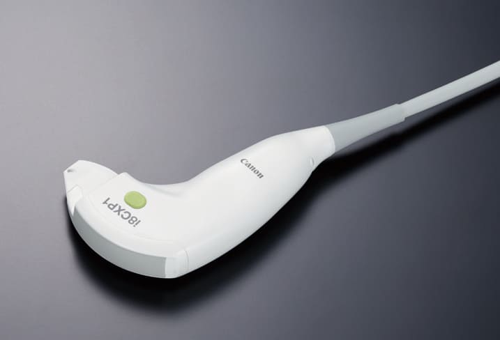
Figure 2 PVI-450BXP transducer
The dedicated interventional transducer, PVI-450BXP (Fig. 2), features a sophisticated design that eliminates potential blind spots in the needle path, ensuring the needle entry within the image field. This transducer is made of single crystal material, providing the high level of image quality essential for safely performing interventions such as Percutaneous Transhepatic Biliary Drainage (PTBD) and Percutaneous Transhepatic Gallbladder Aspiration (PTGBA). Additionally, a disposable attachment is available for urgent cases and repeated procedures.
Improvements in Doppler technology add significant clinical value to ultrasound imaging. SMI is an advanced Doppler technique that enhances the visualization of low flow in the small vessels, minimizing motion artifacts. This technique provides crucial information on tissue blood flow signals, allowing real-time evaluation. Due to the limited vascular phase, timing can be challenging with other modalities, including Contrast-Enhanced Ultrasound (CEUS). However, SMI offers real-time, detailed, reproducible evaluation without contrast agents.
Case 1
A female patient in her 80s presented with a large pancreatic cyst measuring over 20 cm and containing approximately 4,000 cc of fluid. Due to abdominal distention and urinary tract obstruction, abdominal ultrasound was performed for percutaneous drainage. Surgical intervention was not considered suitable due to her advanced age and activities of daily living (ADL). The lesion was assessed using the convex probe, PVI-475BX before the procedure. The 3-HI showed anechoic, clear black contents within the cystic lesion (Fig. 3a), whereas the 2nd harmonic imaging showed a high echoic area (Fig. 3b), posing a challenge in differentiating between noise or debris. However, the 3-HI showed clear wall definition, even in the far field, demonstrating effective performance in assessing the cystic lesion. The drained fluid consisted of a clear serous component. Before the puncture, the linear probe PLI-705BX was utilized to locate the intestinal tract accurately, ensuring a safe procedure. The pig-tail catheter was clearly visualized throughout the procedure by using the biopsy probe PVI-450BXP (Fig. 3c). Additionally, the patient had multiple liver cysts, some of which were complicated cysts. The 3-HI distinctly differentiated these cysts (Fig. 3d), whereas the 2nd harmonic imaging showed artifacts even within the simple cysts (Fig. 3e).
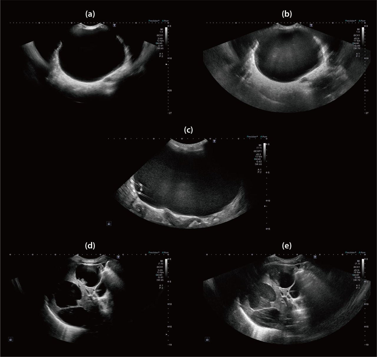
Figure 3 (a) Large cyst image with 3-HI
(b) Large cyst image with 2nd harmonic imaging
(c) Pig-tail catheter can be seen
(d) Liver cyst image with 3-HI
(e) Liver cyst image with 2nd harmonic imaging
Case 2
A male patient in his 20s was referred to our hospital for further investigation and potential surgery after a broadbased gallbladder polyp was detected during a general health check-up. The 2nd harmonic imaging showed broadbased polyps over 1 cm in size, some demonstrating mobility on repositioning, and gallbladder stones were also observed (Figs. 4a and 4b). However, the use of 3-HI enabled a more distinct observation of a stalked polyp within the sludge (Figs. 4c and 4d). Following these findings, the patient was confidently placed under follow-up observation. Other planes showed significant differences between the 3-HI and the 2nd harmonic imaging.
Assessing the gallbladder wall is often challenging. However, the 3-HI offers a clear and defined visualization of Rokitansky-Aschoff Sinus (RAS) with high resolution and high contrast image quality.
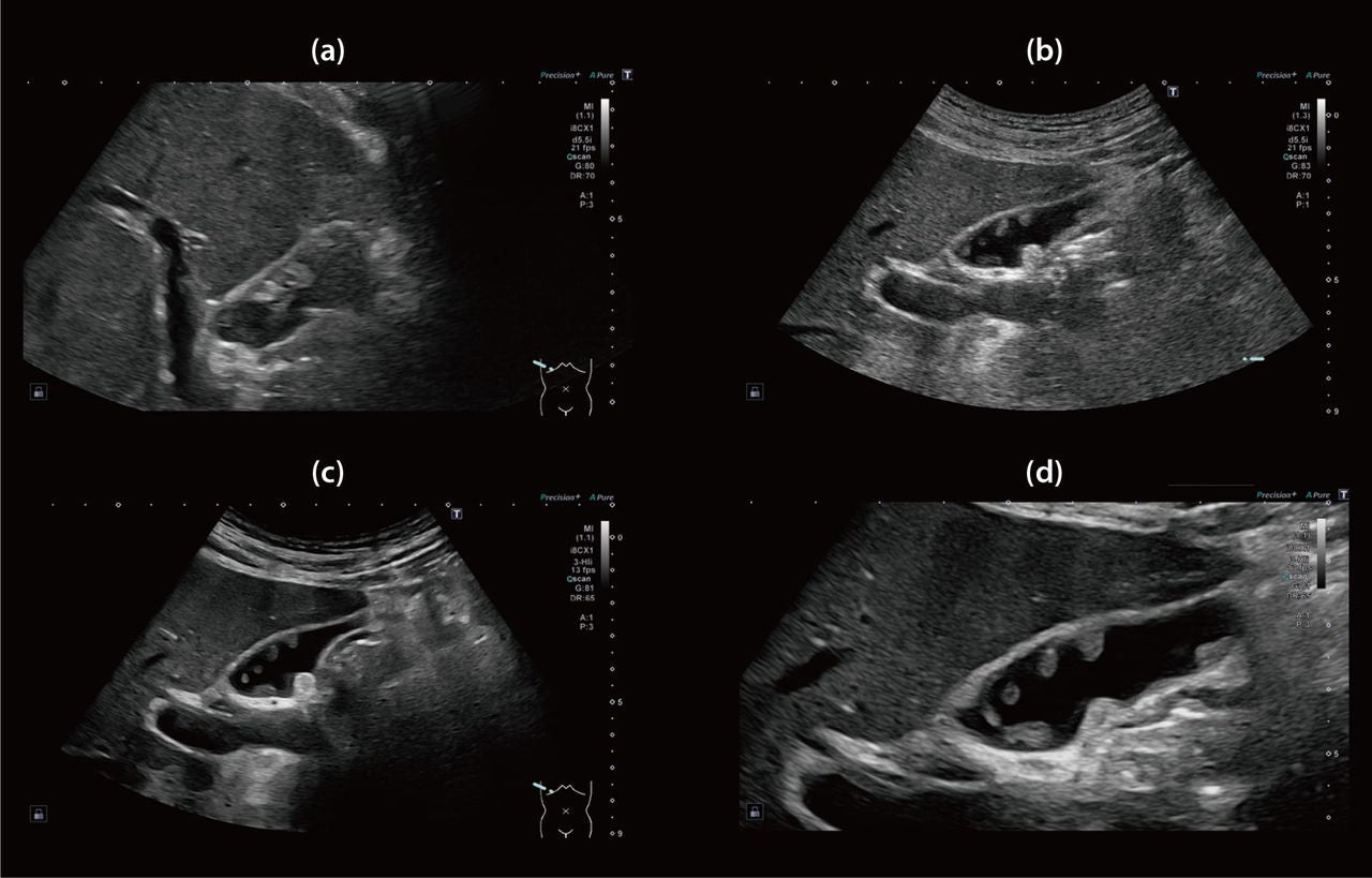
Figure 4 (a)(b) Gallbladder polyp and stone images with 2nd harmonic imaging
(c)(d) Gallbladder polyp and stone images with 3-HI
Case 3
This case involved a male patient with Intraductal Papillary Mucinous Neoplasm (IPMN) who returned for a routine follow-up (Fig. 5a). Localized wall thickening was observed in the septal wall. SMI revealed internal blood flow, indicating hypervascularization (Fig. 5b), and raised suspicions of malignant transformation. Contrast-enhanced CT/EUS also confirmed wall thickening and increased blood flow, leading to the patient undergoing surgical resection.
In a second case, a patient with IPMN under follow-up was examined. The 2nd harmonic imaging detected a small structure within the cyst (Fig. 5c); however, its components were difficult to assess with conventional harmonic imaging. The 3-HI clearly revealed a cystic structure (Fig. 5d), but SMI did not detect any signal.
High image quality with B-mode and SMI provides greater diagnostic confidence in cases like these, where IPMN is a risk factor for pancreatic cancer. Regular follow-up is essential for IPMN management. Transabdominal ultrasound, which can be used repeatedly, is beneficial in these scenarios. Notably, the 3-HI significantly enhances the management and diagnostic accuracy of IPMN patients.
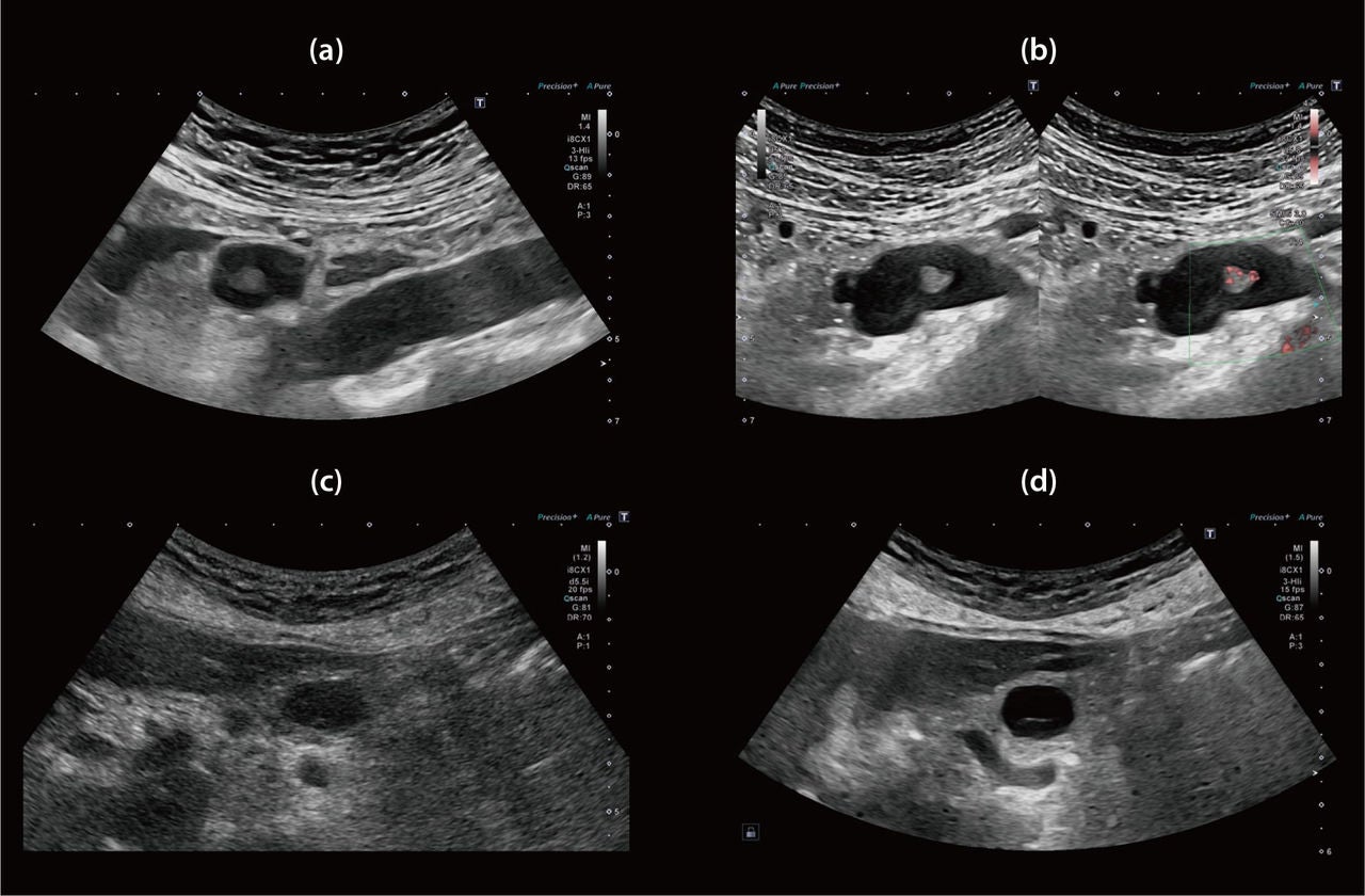
Figure 5 (a) Solid components were observed inside of IPMN
(b) These components had vascularity
(c) Structures were observed inside of IPMN but difficult to assess
(d) 3-HI showed the structure clearly and defined it as a mucinous component
Case 4
A female patient experienced recurrent cholangitis following a choledochojejunostomy. Magnetic Resonance Cholangiopancreatography (MRCP) revealed a defect at the root of the posterior segmental branch. Conventional 2nd harmonic imaging failed to clearly visualize the bile duct (Fig. 6a). In contrast, the 3-HI provided a clear and patent view of the bile duct (Fig. 6b). Pneumobilia was also observed, and the patient underwent follow-up.
In other cases, the 3-HI facilitated easier evaluation of the Intrahepatic Bile Duct (IHBD) due to improved delineation of the bile duct lumen and the wall. The 3-HI (Fig. 6c) demonstrated superior image quality in assessing these cases compared to the 2nd harmonic imaging (Fig. 6d). Ultrasound, with its real-time capability, proved more effective than MRI/ CT in observing pneumobilia. Gas from the jejunum was seen flowing into the IHBD via the anastomosis, as clearly shown in Figure 6e. Tracking the influx of gastrointestinal gas at the anastomosis between the bile duct and jejunum is crucial for confirming pneumobilia.
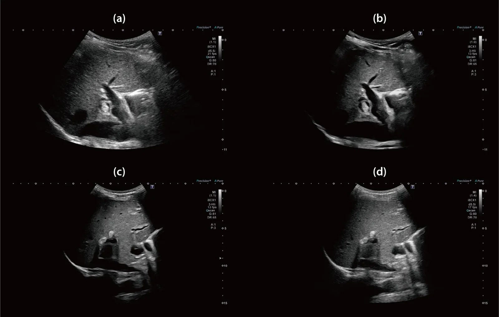
(e)
Figure 6 (a) 2nd harmonic imaging showed a highly echoic lesion in IHBD, but it was difficult to check the obstruction
(b) 3-HI showed IHBD wall and lumen very clearly and confirmed it was not an obstruction
(c) 3-HI showed a better IHBD image compared to (d) 2nd harmonic imaging
(e) Assessment of the stricture at the anastomosis
Case 5
A female patient with a history of metal stent placement for malignant biliary obstruction due to pancreatic cancer presented with fever and abdominal pain. PTGBA was performed to treat cholecystitis caused by blockage of the cystic duct by the metal stent. The design of the PVI-450BXP eliminates any blind spots for the needle at the surface (Fig. 7a). The aspiration was successful (Fig. 7b), and monitoring for bleeding at the end of the procedure was easy (Fig. 7c). Despite the patient’s cachexia, the PVI-450BXP facilitated safe and easy puncturing through the liver. The needle tip was easily identifiable with high image quality, attributed to matrix/single crystal transducer technology.
Young physicians often face limited opportunities to perform procedures due to the paramount importance of patient safety in invasive procedures. However, the challenges can be mitigated with good image quality and a probe design that eliminates blind spots. Additionally, the PVI-450BXP model comes with an available sheath, which is beneficial for infection control. This feature is particularly valuable in pancreatobiliary cases, which frequently require urgent intervention. The availability of a disposable sheath and attachment is highly valued in these scenarios.
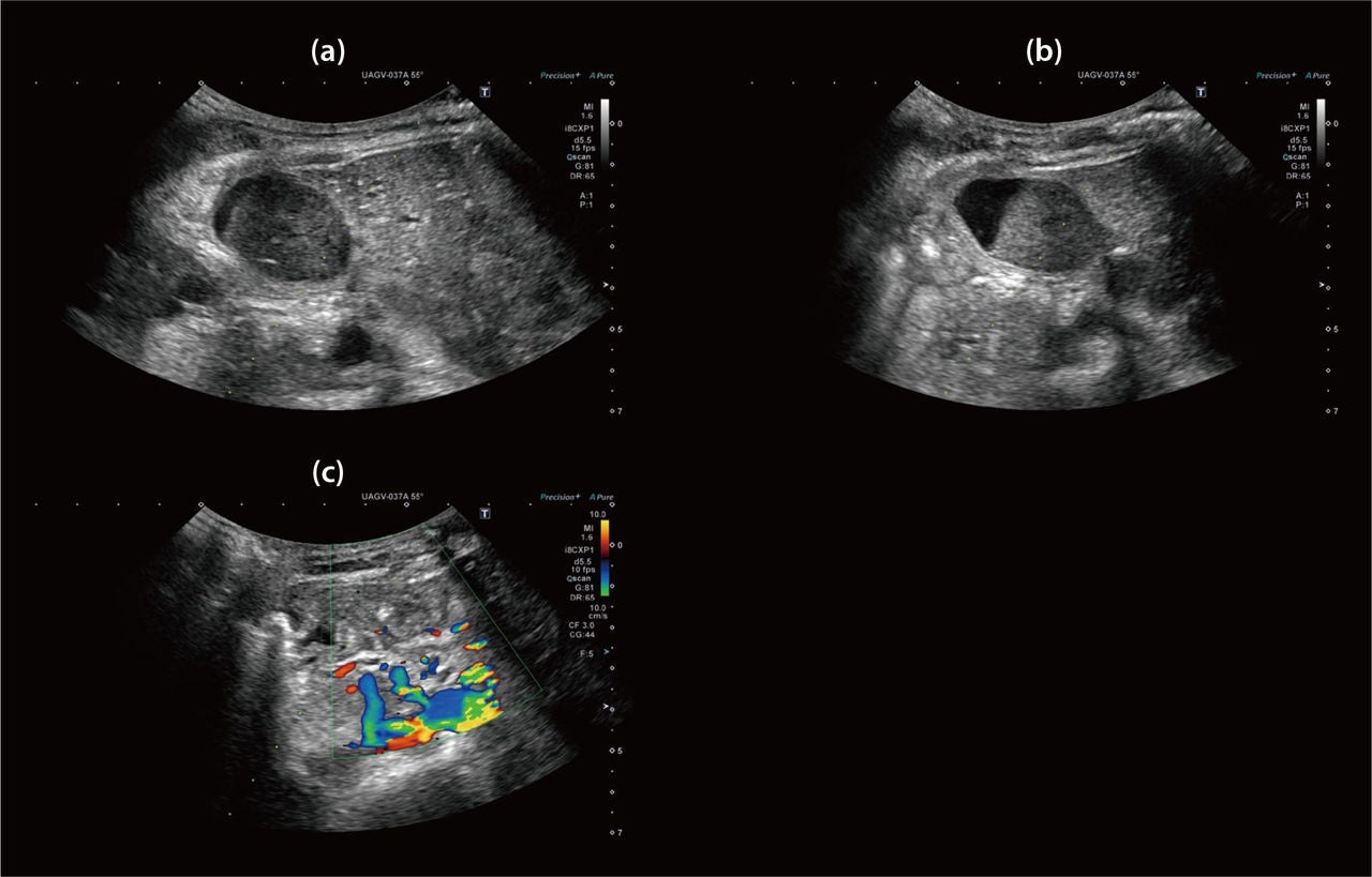
Figure 7 (a) Gallbladder was filled with debris
(b) During the aspiration
(c) Confirmed bleeding
Conclusion
Conventional ultrasound faces a significant challenge in reliably assessing the pancreatobiliary area. The value of 3-HI in the cases discussed is immense, as it has shown a marked improvement in clinical confidence. The numerous advanced technologies integrated into the Aplio i-series / Prism Edition collectively enhance the clinical value for pancreatobiliary physicians. Aplio i-series / Prism Edition offers a comprehensive solution for gastroenterologist.
Social Media


